

Chromosome Synapsis and Recombination in Male-Sterile and Female-Fertile Interspecies Hybrids of the Dwarf Hamsters (Phodopus, Cricetidae)
- PMID: 29693587
- PMCID: PMC5977167
- DOI: 10.3390/genes9050227
Chromosome Synapsis and Recombination in Male-Sterile and Female-Fertile Interspecies Hybrids of the Dwarf Hamsters (Phodopus, Cricetidae)
Abstract
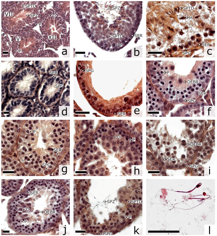
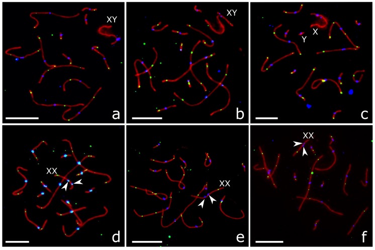
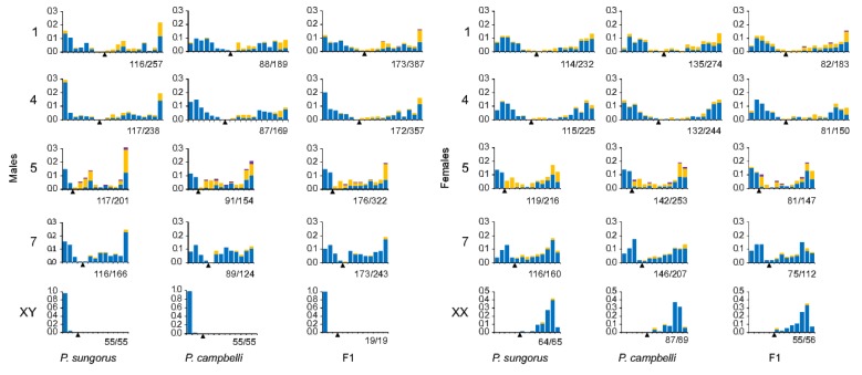
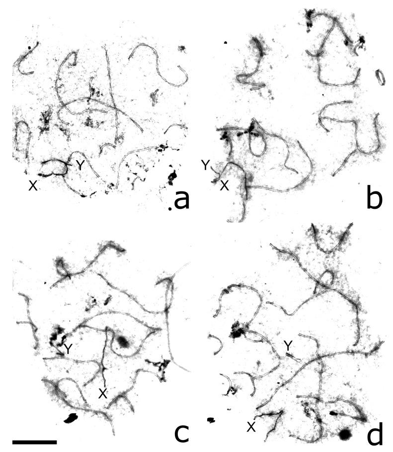
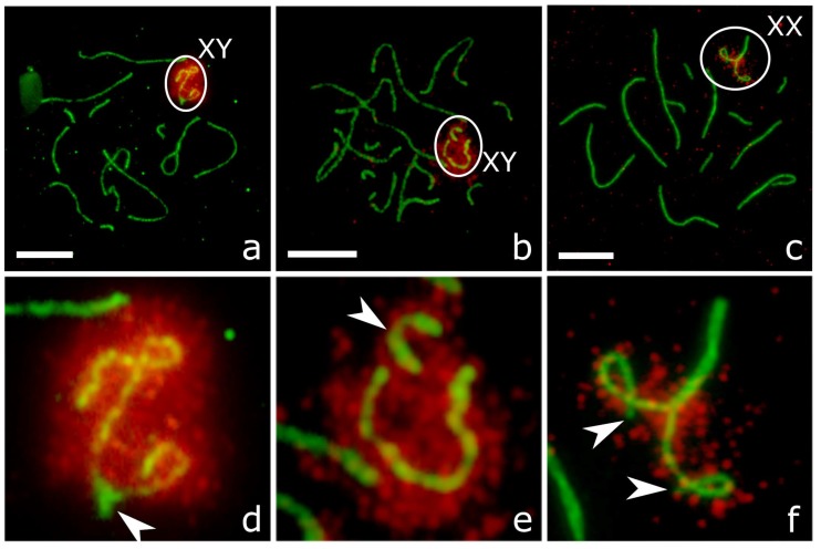
Hybrid sterility is an important step in the speciation process. Hybrids between dwarf hamsters Phodopus sungorus and P.campbelli provide a good model for studies in cytological and genetic mechanisms of hybrid sterility. Previous studies in hybrids detected multiple abnormalities of spermatogenesis and a high frequency of dissociation between the X and Y chromosomes at the meiotic prophase. In this study, we found that the autosomes of the hybrid males and females underwent paring and recombination as normally as their parental forms did. The male hybrids showed a significantly higher frequency of asynapsis and recombination failure between the heterochromatic arms of the X and Y chromosomes than the males of the parental species. Female hybrids as well as the females of the parental species demonstrated a high incidence of centromere misalignment at the XX bivalent and partial asynapsis of the ends of its heterochromatic arms. In all three karyotypes, recombination was completely suppressed in the heterochromatic arm of the X chromosome, where the pseudoautosomal region is located. We propose that this recombination pattern speeds up divergence of the X- and Y-linked pseudoautosomal regions between the parental species and results in their incompatibility in the male hybrids.
Keywords: MLH1; SYCP3; hybrid sterility; pseudoautosomal region; sex chromosomes; synaptonemal complex; γH2A.X.
Conflict of interest statement
The authors declare no conflict of interest. The founding sponsors had no role in the design of the study; in the collection, analyses, or interpretation of data; in the writing of the manuscript, and in the decision to publish the results.
Figures
Similar articles
-
Reproductive Isolation Between Taxonomically Controversial Forms of the Gray Voles (Microtus, Rodentia; Arvicolinae): Cytological Mechanisms and Taxonomical Implications.Front Genet. 2021 May 10;12:653837. doi: 10.3389/fgene.2021.653837. eCollection 2021. Front Genet. 2021. PMID: 34040633 Free PMC article.
-
Abnormal pairing of X and Y sex chromosomes during meiosis I in interspecific hybrids of Phodopus campbelli and P. sungorus.Sci Rep. 2015 Mar 24;5:9435. doi: 10.1038/srep09435. Sci Rep. 2015. PMID: 25801302 Free PMC article.
-
[Characteristics of the first meiotic division in hamster hybrids obtained by backcrossing Phodopus sungorus and Phodopus campbelli].Genetika. 1999 Feb;35(2):237-42. Genetika. 1999. PMID: 10495939 Russian.
-
Interspecific hybrids of dwarf hamsters and Phasianidae birds as animal models for studying the genetic and developmental basis of hybrid incompatibility.Genes Genet Syst. 2016 Oct 13;91(2):63-75. doi: 10.1266/ggs.16-00022. Epub 2016 Sep 15. Genes Genet Syst. 2016. PMID: 27628130 Review.
-
Misregulation of Gene Expression and Sterility in Interspecies Hybrids: Causal Links and Alternative Hypotheses.J Mol Evol. 2016 May;82(4-5):176-82. doi: 10.1007/s00239-016-9734-z. Epub 2016 Mar 29. J Mol Evol. 2016. PMID: 27025762 Review.
Cited by
-
CENP-A binding domains and recombination patterns in horse spermatocytes.Sci Rep. 2019 Nov 1;9(1):15800. doi: 10.1038/s41598-019-52153-1. Sci Rep. 2019. PMID: 31676881 Free PMC article.
-
Different complex regulatory phenotypes underlie hybrid male sterility in divergent rodent crosses.bioRxiv [Preprint]. 2024 Nov 11:2023.10.30.564782. doi: 10.1101/2023.10.30.564782. bioRxiv. 2024. Update in: Genetics. 2025 Feb 05;229(2):iyae198. doi: 10.1093/genetics/iyae198. PMID: 37961317 Free PMC article. Updated. Preprint.
-
Reproductive Isolation Between Taxonomically Controversial Forms of the Gray Voles (Microtus, Rodentia; Arvicolinae): Cytological Mechanisms and Taxonomical Implications.Front Genet. 2021 May 10;12:653837. doi: 10.3389/fgene.2021.653837. eCollection 2021. Front Genet. 2021. PMID: 34040633 Free PMC article.
-
Relationship between meiotic behaviour and fertility in backcross-1 derivatives of the [(Gossypium hirsutum × G. thurberi)2 × G. longicalyx] trispecies hybrid.Comp Cytogenet. 2020 Jan 28;14(1):75-95. doi: 10.3897/CompCytogen.v14i1.47231. eCollection 2020. Comp Cytogenet. 2020. PMID: 32047586 Free PMC article.
-
Different complex regulatory phenotypes underlie hybrid male sterility in divergent rodent crosses.Genetics. 2025 Feb 5;229(2):iyae198. doi: 10.1093/genetics/iyae198. Genetics. 2025. PMID: 39601270 Free PMC article.
References
-
- Coyne J.A., Orr H.A. Speciation. Sinauer Associates; Sunderland, MA, USA: 2004.
-
- Benirschke K. Comparative Aspects of Reproductive Failure. Springer; Berlin/Heidelberg, Germany: 1967. Sterility and Fertility of Interspecific Mammalian Hybrids; pp. 218–234.
LinkOut - more resources
Full Text Sources
Other Literature Sources
