

Comparative proteomic analyses of human adipose extracellular matrices decellularized using alternative procedures
- PMID: 29693792
- PMCID: PMC6158104
- DOI: 10.1002/jbm.a.36444
Comparative proteomic analyses of human adipose extracellular matrices decellularized using alternative procedures
Abstract
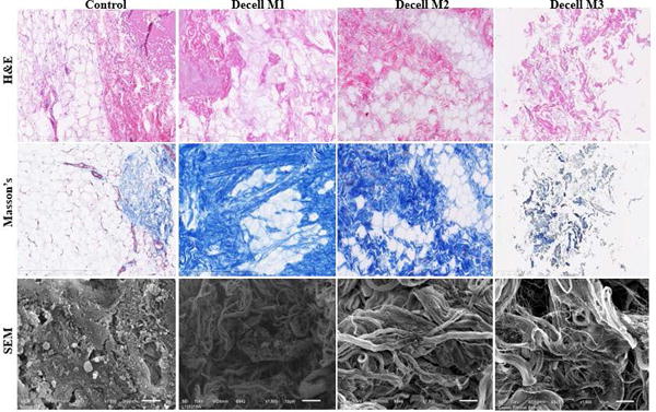
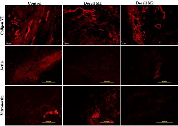
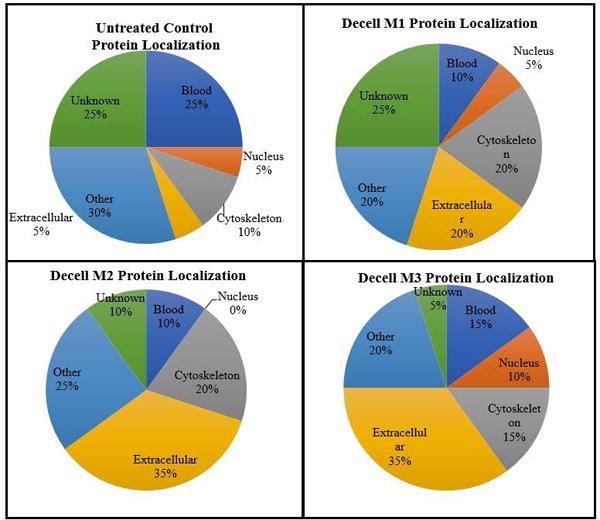
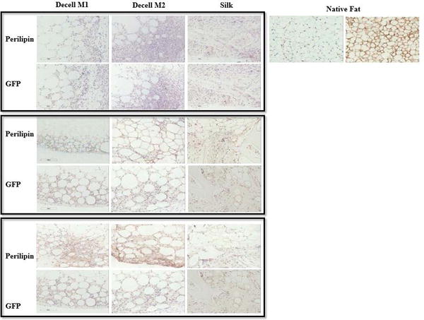
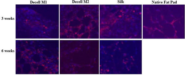
Decellularized human adipose tissue has potential clinical utility as a processed biological scaffold for soft tissue cosmesis, grafting, and reconstruction. Adipose tissue decellularization has been accomplished using enzymatic-, detergent-, and/or solvent-based methods. To examine the hypothesis that distinct decellularization processes may yield scaffolds with differing compositions, the current study employed mass spectrometry to compare the proteomes of human adipose-derived matrices generated through three independent methods combining enzymatic-, detergent-, and/or solvent-based steps. In addition to protein content, bioscaffolds were evaluated for deoxyribose nucleic acid depletion, extracellular matrix composition, and physical structure using optical density, histochemical staining, and scanning electron microscopy. Mass spectrometry based proteomic analyses identified 25 proteins (having at least two peptide sequences detected) in the scaffolds generated with an enzymatic approach, 143 with the detergent approach, and 102 with the solvent approach, as compared to 155 detected in unprocessed native human fat. Immunohistochemical detection confirmed the presence of the structural proteins actin, collagen type VI, fibrillin, laminin, and vimentin. Subsequent in vivo analysis of the predominantly enzymatic- and detergent-based decellularized scaffolds following subcutaneous implantation in GFP+ transgenic mice demonstrated that the matrices generated with both approaches supported the ingrowth of host-derived adipocyte progenitors and vasculature in a time dependent manner. Together, these results determine that decellularization methods influence the protein composition of adipose tissue-derived bioscaffolds. © 2018 Wiley Periodicals, Inc. J Biomed Mater Res Part A: 106A:2481-2493, 2018.
Keywords: adipose tissue; bioscaffold; decellularization; extracellular matrix; mass spectrometry proteomics; regenerative medicine.
© 2018 Wiley Periodicals, Inc.
Figures
References
-
- Kaufman MR, Bradley JP, Dickinson B, Heller JB, Wasson K, O’Hara C, Huang C, Gabbay J, Ghadjar K, Miller TA. Autologous fat transfer national consensus survey: trends in techniques for harvest, preparation, and application, and perception of short- and long-term results. Plastic and reconstructive surgery. 2007;119(1):323–31. - PubMed
-
- Coudurier J, Ho Quoc C, Ismail M, Dlimi C, Tourasse C, Delay E. Long-term outcome of lipomodeling in Poland’s syndrome: about our first case with an eleven-years’ follow-up. Ann Chir Plast Esthet. 2015;60(1):65–9. - PubMed
-
- Marra KG, Rubin JP. The potential of adipose-derived stem cells in craniofacial repair and regeneration. Birth Defects Res C Embryo Today. 2012;96(1):95–7. - PubMed
-
- Yoshimura K, Sato K, Aoi N, Kurita M, Inoue K, Suga H, Eto H, Kato H, Hirohi T, Harii K. Cell-assisted lipotransfer for facial lipoatrophy: efficacy of clinical use of adipose-derived stem cells. Dermatologic surgery: official publication for American Society for Dermatologic Surgery [et al] 2008;34(9):1178–85. - PubMed
-
- Pallua N, Baroncini A, Alharbi Z, Stromps JP. Improvement of facial scar appearance and microcirculation by autologous lipofilling. Journal of plastic, reconstructive & aesthetic surgery: JPRAS. 2014;67(8):1033–7. - PubMed
Publication types
MeSH terms
Substances
Grants and funding
LinkOut - more resources
Full Text Sources
Other Literature Sources
