Wistar rats immature testicular tissue vitrification and heterotopic grafting
- PMID: 29693963
- PMCID: PMC6106629
- DOI: 10.5935/1518-0557.20180023
Wistar rats immature testicular tissue vitrification and heterotopic grafting
Abstract
Objective: To evaluate the efficiency of two vitrification protocols for rat immature testicular tissue and heterotopic transplantation.
Methods: Twenty-four pre-pubertal Wistar rats were divided into three groups (n=8). After orchiectomy, testicular fragments (3mm) from Groups 1 and 2 were vitrified with different cryoprotectant concentration solutions, using sterile inoculation loops as support. After warming up, the fragments were submitted to cell viability assessment by Trypan blue and histological evaluation. Vitrified (Groups 1 and 2) and fresh (Group 3) fragments were grafted to the animals periauricular region. After 8 weeks of grafting, the implant site was histologically analyzed.
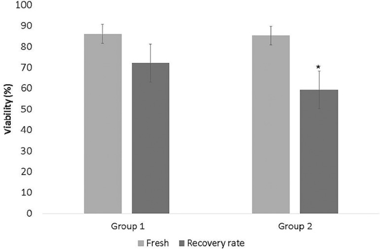
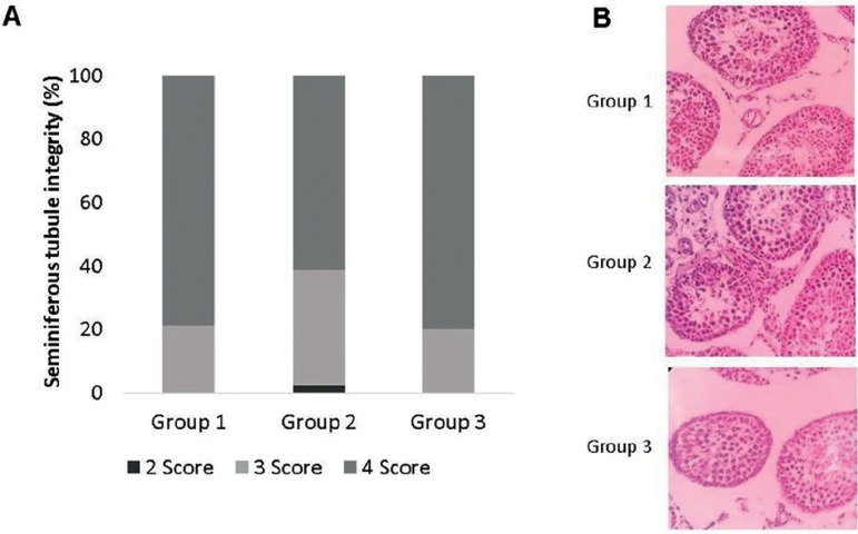
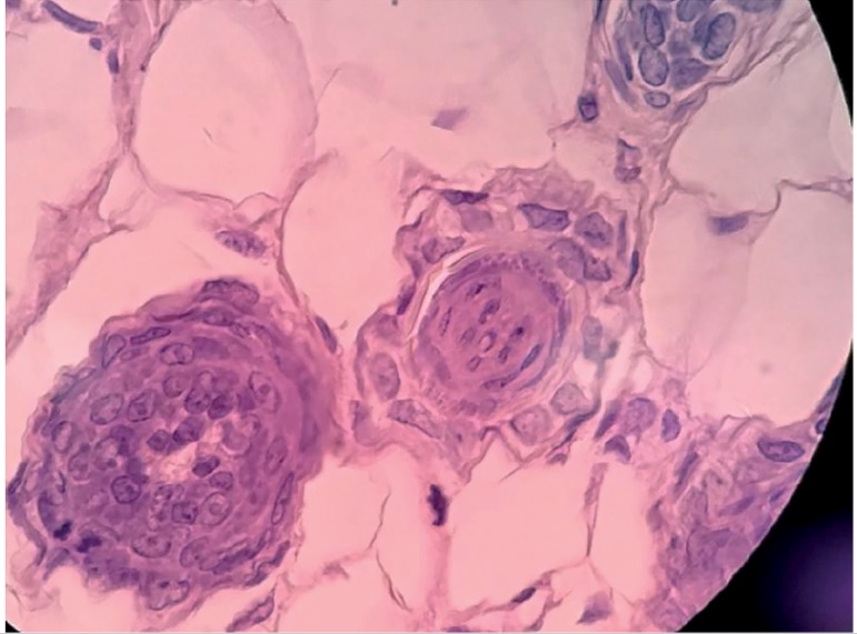
Results: The viability recovery rate from Group 1 (72.09%) was higher (p=0.02) than that from Group 2 (59.19%). Histological analysis showed similar tubular integrity between fresh fragments from Groups 1 and 3. Group 2 samples presented lower tubular integrity. We ran histological analyses in the grafts from the Groups. In all groups, it was possible to see the implant site, however, no fragment of testicular tissue or signs of inflammation were histologically found in most samples from Groups 1 and 3. In one sample from Group 2, we found degenerated seminiferous tubules with necrosis and signs of an inflammatory process. In another sample from Group 2, we found seminiferous tubules in the implant site.
Conclusion: The vitrification of pre-pubertal testicular tissue of rats showed little damage to cell viability through histological analysis when we used cryoprotectants in a lower concentration. Heterotopic transplantation could not preserve the structural organization of the testicular tissue.
Keywords: cryopreservation; fertility preservation; prepubertal; transplantation.
Figures

References
-
- Borges Júnior E, Braga DPAF, Radaelli MRM. Criopreservação de tecido germinativo masculino. In: Almodin CG, Costa RR, editors. Criopreservação em reprodução. Maringá: Dental Press; 2014. pp. 169–194.
MeSH terms
LinkOut - more resources
Full Text Sources
Other Literature Sources
Miscellaneous