. 2018 Apr 25;13(4):e0196455.
doi: 10.1371/journal.pone.0196455. eCollection 2018.

Haptoglobin and hemopexin inhibit vaso-occlusion and inflammation in murine sickle cell disease: Role of heme oxygenase-1 induction

Affiliations

Haptoglobin and hemopexin inhibit vaso-occlusion and inflammation in murine sickle cell disease: Role of heme oxygenase-1 induction

John D Belcher et al. PLoS One. .

Abstract

During hemolysis, hemoglobin and heme released from red blood cells promote oxidative stress, inflammation and thrombosis. Plasma haptoglobin and hemopexin scavenge free hemoglobin and heme, respectively, but can be depleted in hemolytic states. Haptoglobin and hemopexin supplementation protect tissues, including the vasculature, liver and kidneys. It is widely assumed that these protective effects are due primarily to hemoglobin and heme clearance from the vasculature. However, this simple assumption does not account for the consequent cytoprotective adaptation seen in cells and organs. To further address the mechanism, we used a hyperhemolytic murine model (Townes-SS) of sickle cell disease to examine cellular responses to haptoglobin and hemopexin supplementation. A single infusion of haptoglobin or hemopexin (± equimolar hemoglobin) in SS-mice increased heme oxygenase-1 (HO-1) in the liver, kidney and skin several fold within 1 hour and decreased nuclear NF-ĸB phospho-p65, and vaso-occlusion for 48 hours after infusion. Plasma hemoglobin and heme levels were not significantly changed 1 hour after infusion of haptoglobin or hemopexin. Haptoglobin and hemopexin also inhibited hypoxia/reoxygenation and lipopolysaccharide-induced vaso-occlusion in SS-mice. Inhibition of HO-1 activity with tin protoporphyrin blocked the protections afforded by haptoglobin and hemopexin in SS-mice. The HO-1 reaction product carbon monoxide, fully restored the protection, in part by inhibiting Weibel-Palade body mobilization of P-selectin and von Willebrand factor to endothelial cell surfaces. Thus, the mechanism by which haptoglobin and hemopexin supplementation in hyperhemolytic SS-mice induces cytoprotective cellular responses is linked to increased HO-1 activity.


Conflict of interest statement

Competing Interests: CSL Behring and their employees [SMM NB] are developing haptoglobin and hemopexin for commercial use. The commercial affiliation does not alter our adherence to PLOS ONE policies on sharing data and materials. The specific roles of these authors are articulated with their initials in the 'author contributions ' section.

Figures

Fig 1
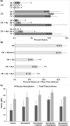
Fig 1. Haptoglobin and hemopexin inhibit hemoglobin-induced stasis in SS-mice.
Dorsal skin-fold chambers were implanted onto Townes-SS mice (n = 4/group) and Townes-AA mice (n = 3/group) and 20–24 flowing venules were selected in each mouse at baseline (time 0). (A) SS and AA-mice were untreated or SS-mice were infused with haptoglobin (Hp, 1 μmol/kg) or hemopexin (Hpx, 1 μmol/kg) at baseline after selection of flowing venules. Microvascular stasis (% non-flowing venules) was measured in the same venules at 24, 48 and 72 hours. Bars represent means ± SD. *P < .05 versus SS (untreated). (B) SS-mice with implanted dorsal skin-fold chambers (n = 3/group) were infused with equimolar concentrations (1 μmol/kg) of hemoglobin (Hb), Hb + albumin, Hb + Hp, Hb + Hpx, or Hb + Hp + Hpx (0.5 μmol/kg each of Hp and Hpx). Microvascular stasis was measured 1 hour after infusion. Values are means ± SD. **P ≤ .01 versus Hb and Hb + albumin. (C) SS-mice without dorsal skin-fold chambers (n = 4/group) were infused with vehicle (saline), Hb, Hb + albumin, Hb + Hp, or Hb + Hpx at equimolar concentrations (1 μmol/kg). Total plasma heme and Hb levels were measured in venous plasma samples collected 1 hour after infusion. Bars are means ± SD.
Fig 2
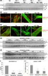
Fig 2. Haptoglobin and hemopexin inhibit inflammatory markers.
(A) SS-mice (n = 3/group) were infused with equimolar concentrations (1 μmol/kg) of Hb, Hb + albumin, Hb + Hp, Hb + Hpx, or Hb + Hp + Hpx. Livers were removed and flash frozen 4 hours after infusion. NF-κB phospho- and total p65 expression was assessed in hepatic nuclear extracts by immunoblot. (B and C) ICAM-1 (red) or VCAM-1 (red) and CD31 (green) immunofluorescence in dorsal skin samples from SS-mice 4 hours after infusion of vehicle, Hb, Hb + Hp, or Hb + Hpx. (D and E) SS-mice (n = 3/group) were infused with vehicle or increasing doses (0.0156, 0.0625, 0.25 or 1.0 μmols/kg) of Hp or Hpx at baseline. Livers were removed and flash frozen 24 hours after infusion. NF-κB phospho- and total p65 expression was assessed in hepatic nuclear extracts by immunoblot. (F) RANTES/CCL5 levels in the plasma of SS-mice (n = 3/group) 24 hours after infusion of vehicle, Hp or Hpx (1 μmol/kg). Bars are means ± SD, *p < .01 versus vehicle). (G) 4-Hydroxynonenal (4-HNE) levels in liver microsomes of SS-mice (n = 3/group) 24 hours after infusion of vehicle, Hp or Hpx (1 μmol/kg). Bars are means ± SD, *p < .01 versus vehicle.
Fig 3
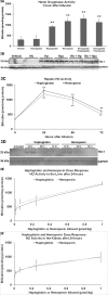
Fig 3. HO-1 is rapidly increased after haptoglobin and hemopexin infusion.
(A and B) SS-mice (n = 3/group) were infused with vehicle or equimolar (1 μmol/kg) Hb, Hp, Hpx, Hb + Hp, or Hb + Hpx. Livers were removed and flash frozen 1 hour after infusion. Hepatic microsomes were used to assess heme oxygenase (HO) activity (A) via bilirubin production and protein expression (B) via immunoblot. Bars are means ± SD, **p < .01 versus vehicle or Hb. (C and D) SS-mice (n = 3/group) were untreated or infused with Hp or Hpx (1 μmol/kg) at baseline (time 0). Livers were removed and flash frozen 24, 48 or 72 hours after infusion. Hepatic microsomes were used to assess (C) HO activity and (D) HO-1 protein expression via immunoblot. Bars are means ± SD, *p < .05 and **p < .01 versus untreated SS-mice. (E and F) SS-mice (n = 3/group) were infused with vehicle or increasing doses (0.0156, 0.0625, 0.25 or 1.0 μmols/kg) of Hp or Hpx at baseline. Livers and kidneys were removed and flash frozen 24 hours after infusion. Hepatic (E) and kidney (F) microsomes were used to assess HO activity. Bars are means ± SD.
Fig 4
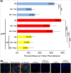
Fig 4. The HO-1 inhibitor tin protoporphyrin (SnPP) blocks the inhibition of stasis by haptoglobin and hemopexin and is reversed by carbon monoxide (CO).
(A) Three groups of SS-mice (n = 9/group) with implanted dorsal skin-fold chambers were infused with equimolar (1 μmol/kg) Hb (n = 3), Hb + Hp (n = 3), or Hb + Hpx (n = 3). The first group (blue bars) had no pretreatments prior to infusion. The second group (red bars) was pretreated with the HO inhibitor SnPP (40 μmol/kg i.p. X 3 days) prior to infusion. The third group (yellow bars) was pretreated with SnPP and inhaled CO (250 ppm in air X 1h/day X 3 days) prior to infusion. Immediately prior to infusion, 20–24 flowing venules were selected in each mouse. Microvascular stasis was measured in the same venules 1 hour after infusion. Bars represent means ± SD. **P < .01. (B) Human umbilical vein endothelial cells (HUVEC) were incubated ± 80 μM CO-releasing molecule (CORM) 1A or CORM 2 for 30 minutes followed by treatment with 10 μM hemin for 30 minutes. HUVEC treated with 100 μM histamine served as a positive control for Weibel-Palade body P-selectin and VWF expression on the cell surface. Green and red fluorescence denote P-selectin and VWF expression, respectively, on the surface of HUVEC. The blue fluorescence denotes nuclei. Magnification is 60X. White bars in images represent 10 μm.
Fig 5
Fig 5. Haptoglobin and hemopexin inhibit stasis in SS-mice challenged with hypoxia-reoxygenation (H/R) and lipopolysaccharide (LPS).
Dorsal skin-fold chambers were implanted onto SS-mice (n = 3/group) and 20–25 flowing venules were selected in each mouse. After venule selection, mice were infused with vehicle, Hp (1 μmol/kg), Hpx (1 μmol/kg), or Hp + Hpx (0.5 μmol/kg each of Hp and Hpx). The Hp + Hpx mice were pretreated with SnPP (40 μmols/kg i.p. X 3 days). One hour after infusion, mice were challenged with H/R (7% O2 for 1h followed by room air for 1h) or LPS (1 mg/kg, i.p.). Microvascular stasis was measured after H/R or one hour after LPS administration. Bars are means ± SD. **P < .01.
Fig 6
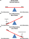
Fig 6. Tipping point: A delicate balance between pro-inflammatory and anti-inflammatory forces during steady-state sickle cell disease.
Based on our data we propose a model of vaso-occlusion (stasis) in SCD where in steady-state pro-inflammatory heme and anti-inflammatory HO-1 are in a delicate balance (A). Because of this delicate balance a relatively small increase in hemolysis or plasma hemoglobin/heme can tip the balance in favor of a pro-inflammatory state and more stasis (B). And conversely, increasing HO-1 expression by administration of haptoglobin or hemopexin or other methods tips the balance in favor of an anti-inflammatory state and less stasis (C).

References

    1. Reiter CD, Wang X, Tanus-Santos JE, Hogg N, Cannon RO 3rd, Schechter AN, et al. Cell-free hemoglobin limits nitric oxide bioavailability in sickle-cell disease. Nat Med. 2002;8(12):1383–9. Epub 2002/11/12. doi: 10.1038/nm799 . - DOI - PubMed
    1. Cooper CE, Silaghi-Dumitrescu R, Rukengwa M, Alayash AI, Buehler PW. Peroxidase activity of hemoglobin towards ascorbate and urate: a synergistic protective strategy against toxicity of Hemoglobin-Based Oxygen Carriers (HBOC). Biochim Biophys Acta. 2008;1784(10):1415–20. Epub 2008/05/07. doi: 10.1016/j.bbapap.2008.03.019 . - DOI - PubMed
    1. Chintagari NR, Jana S, Alayash AI. Oxidized Ferric and Ferryl Forms of Hemoglobin Trigger Mitochondrial Dysfunction and Injury in Alveolar Type I Cells. Am J Respir Cell Mol Biol. 2016;55(2):288–98. Epub 2016/03/15. doi: 10.1165/rcmb.2015-0197OC ; PubMed Central PMCID: PMCPMC4979363. - DOI - PMC - PubMed
    1. Bunn HF, Jandl JH. Exchange of heme among hemoglobins and between hemoglobin and albumin. J Biol Chem. 1968;243(3):465–75. Epub 1968/02/10. . - PubMed
    1. Belcher JD, Chen C, Nguyen J, Milbauer L, Abdulla F, Alayash AI, et al. Heme triggers TLR4 signaling leading to endothelial cell activation and vaso-occlusion in murine sickle cell disease. Blood. 2014;123(3):377–90. doi: 10.1182/blood-2013-04-495887 . - DOI - PMC - PubMed

Publication types

MeSH terms