

Long non-coding RNA-ROR aggravates myocardial ischemia/reperfusion injury
- PMID: 29694511
- PMCID: PMC5937723
- DOI: 10.1590/1414-431x20186555
Long non-coding RNA-ROR aggravates myocardial ischemia/reperfusion injury
Retraction in
-
Retraction notice for: "Long non-coding RNA-ROR aggravates myocardial ischemia/reperfusion injury" [Braz J Med Biol Res (2018) 51(6): e6555].Braz J Med Biol Res. 2021 Jun 2;54(6):e6555retraction. doi: 10.1590/1414-431X2020e6555retraction. Braz J Med Biol Res. 2021. PMID: 34133474 Free PMC article.
Abstract
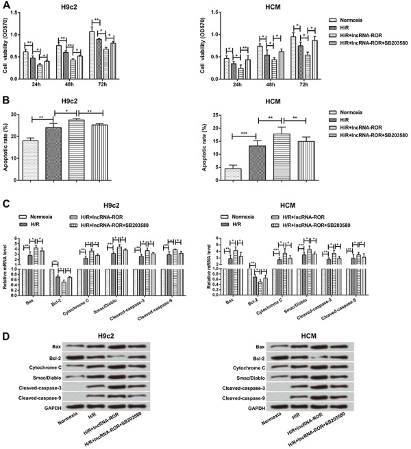
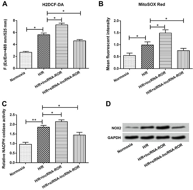
Long non-coding RNAs (lncRNAs) play an important role in the pathogenesis of cardiovascular diseases, especially in myocardial infarction and ischemia/reperfusion (I/R). However, the underlying molecular mechanism remains unclear. In this study, we determined the role and the possible underlying molecular mechanism of lncRNA-ROR in myocardial I/R injury. H9c2 cells and human cardiomyocytes (HCM) were subjected to either hypoxia/reoxygenation (H/R), I/R or normal conditions (normoxia). The expression levels of lncRNA-ROR were detected in serum of myocardial I/R injury patients, H9c2 cells, and HCM by qRT-PCR. Then, levels of lactate dehydrogenase (LDH), malondialdehyde (MDA), superoxide dismutase (SOD), and glutathione peroxidase (GSH-PX) were measured by kits. Cell viability, apoptosis, apoptosis-associated factors, and p38/MAPK pathway were examined by MTT, flow cytometry, and western blot assays. Furthermore, reactive oxygen species (ROS) production was determined by H2DCF-DA and MitoSOX Red probes with flow cytometry. NADPH oxidase activity and NOX2 protein levels were measured by lucigenin chemiluminescence and western blot. Results showed that lncRNA-ROR expression was increased in I/R patients and in H/R treatment of H9c2 cells and HCM. Moreover, lncRNA-ROR significantly promoted H/R-induced myocardial injury via stimulating release of LDH, MDA, SOD, and GSH-PX. Furthermore, lncRNA-ROR decreased cell viability, increased apoptosis, and regulated expression of apoptosis-associated factors. Additionally, lncRNA-ROR increased phosphorylation of p38 and ERK1/2 expression and inhibition of p38/MAPK, and rescued lncRNA-ROR-induced cell injury in H9c2 cells and HCM. ROS production, NADPH oxidase activity, and NOX2 protein levels were promoted by lncRNA-ROR. These data suggested that lncRNA-ROR acted as a therapeutic agent for the treatment of myocardial I/R injury.
Figures


Similar articles
-
Long non-coding RNA ROR sponges miR-138 to aggravate hypoxia/reoxygenation-induced cardiomyocyte apoptosis via upregulating Mst1.Exp Mol Pathol. 2020 Jun;114:104430. doi: 10.1016/j.yexmp.2020.104430. Epub 2020 Mar 30. Exp Mol Pathol. 2020. PMID: 32240614
-
GAS5 promotes myocardial apoptosis in myocardial ischemia-reperfusion injury via upregulating LAS1 expression.Eur Rev Med Pharmacol Sci. 2018 Dec;22(23):8447-8453. doi: 10.26355/eurrev_201812_16544. Eur Rev Med Pharmacol Sci. 2018. PMID: 30556886
-
MicroRNA-208a Regulates H9c2 Cells Simulated Ischemia-Reperfusion Myocardial Injury via Targeting CHD9 through Notch/NF-kappa B Signal Pathways.Int Heart J. 2018 May 30;59(3):580-588. doi: 10.1536/ihj.17-147. Epub 2018 May 20. Int Heart J. 2018. PMID: 29681568
-
Long noncoding RNAs: Important participants and potential therapeutic targets for myocardial ischaemia reperfusion injury.Clin Exp Pharmacol Physiol. 2020 Nov;47(11):1783-1790. doi: 10.1111/1440-1681.13375. Epub 2020 Aug 11. Clin Exp Pharmacol Physiol. 2020. PMID: 32621522 Review.
-
Non-coding RNAs modulate autophagy in myocardial ischemia-reperfusion injury: a systematic review.J Cardiothorac Surg. 2021 May 22;16(1):140. doi: 10.1186/s13019-021-01524-9. J Cardiothorac Surg. 2021. PMID: 34022925 Free PMC article.
Cited by
-
Long non‑coding RNA GAS5 regulates myocardial ischemia‑reperfusion injury through the PI3K/AKT apoptosis pathway by sponging miR‑532‑5p.Int J Mol Med. 2020 Mar;45(3):858-872. doi: 10.3892/ijmm.2020.4471. Epub 2020 Jan 20. Int J Mol Med. 2020. PMID: 31985016 Free PMC article.
-
LncRNA RNA ROR Aggravates Hypoxia/Reoxygenation-Induced Cardiomyocyte Ferroptosis by Targeting miR-769-5p/CBX7 Axis.Biochem Genet. 2024 Oct;62(5):3586-3604. doi: 10.1007/s10528-023-10587-3. Epub 2023 Dec 29. Biochem Genet. 2024. PMID: 38157079
-
Cytoprotective Effects of Dinitrosyl Iron Complexes on Viability of Human Fibroblasts and Cardiomyocytes.Front Pharmacol. 2019 Nov 11;10:1277. doi: 10.3389/fphar.2019.01277. eCollection 2019. Front Pharmacol. 2019. PMID: 31780929 Free PMC article.
-
The protective effects of Olmesartan against interleukin-29 (IL-29)-induced type 2 collagen degradation in human chondrocytes.Bioengineered. 2022 Jan;13(1):1802-1813. doi: 10.1080/21655979.2021.1997090. Bioengineered. 2022. Retraction in: Bioengineered. 2024 Dec;15(1):2299551. doi: 10.1080/21655979.2024.2299551. PMID: 35012432 Free PMC article. Retracted.
-
Long non-coding RNA TUG1 knockdown promotes autophagy and improves acute renal injury in ischemia-reperfusion-treated rats by binding to microRNA-29 to silence PTEN.BMC Nephrol. 2021 Aug 24;22(1):288. doi: 10.1186/s12882-021-02473-0. BMC Nephrol. 2021. PMID: 34429073 Free PMC article.
References
Publication types
MeSH terms
Substances
LinkOut - more resources
Full Text Sources
Other Literature Sources
Miscellaneous
