. 2018 Apr 24;114(8):2014-2023.
doi: 10.1016/j.bpj.2018.03.004.

Cytoskeleton Remodeling Induces Membrane Stiffness and Stability Changes of Maturing Reticulocytes

Affiliations

Cytoskeleton Remodeling Induces Membrane Stiffness and Stability Changes of Maturing Reticulocytes

He Li et al. Biophys J. .

Abstract

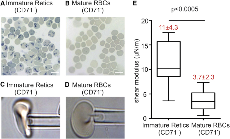
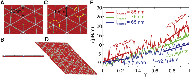
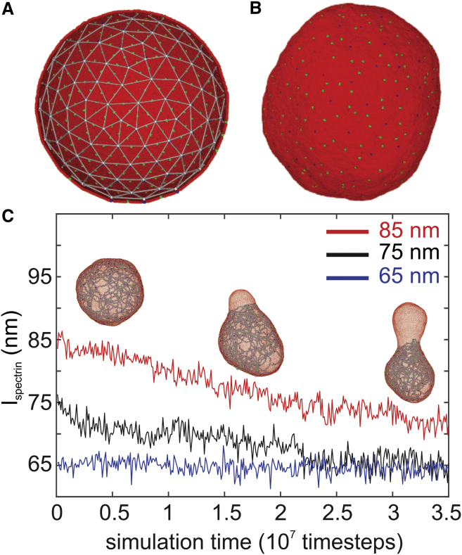
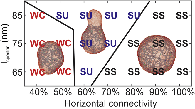
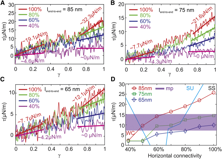
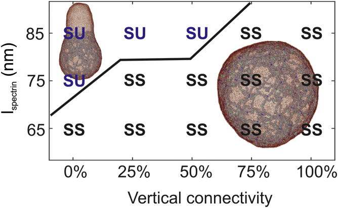
Reticulocytes, the precursors of erythrocytes, undergo drastic alterations in cell size, shape, and deformability during maturation. Experimental evidence suggests that young reticulocytes are stiffer and less stable than their mature counterparts; however, the underlying mechanism is yet to be fully understood. Here, we develop a coarse-grained molecular-dynamics reticulocyte membrane model to elucidate how the membrane structure of reticulocytes contributes to their particular biomechanical properties and pathogenesis in blood diseases. First, we show that the extended cytoskeleton in the reticulocyte membrane is responsible for its increased shear modulus. Subsequently, we quantify the effect of weakened cytoskeleton on the stiffness and stability of reticulocytes, via which we demonstrate that the extended cytoskeleton along with reduced cytoskeleton connectivity leads to the seeming paradox that reticulocytes are stiffer and less stable than the mature erythrocytes. Our simulation results also suggest that membrane budding and the consequent vesiculation of reticulocytes can occur independently of the endocytosis-exocytosis pathway, and thus, it may serve as an additional means of removing unwanted membrane proteins from reticulocytes. Finally, we find that membrane budding is exacerbated when the cohesion between the lipid bilayer and the cytoskeleton is compromised, which is in accord with the clinical observations that erythrocytes start shedding membrane surface at the reticulocyte stage in hereditary spherocytosis. Taken together, our results quantify the stiffness and stability change of reticulocytes during their maturation and provide, to our knowledge, new insights into the pathogenesis of hereditary spherocytosis and malaria.


Figures

Figure 1
Figure 1
Blood smears stained with methylene blue show a reticular staining pattern indicative of residual RNA present in the CD71+ population (A), but not in the CD71 population (B). Scale bars in the figures represent 10 μm. (C) Immature reticulocytes (CD71+) and (D) mature RBCs (CD71) studied by using micropipette aspiration with pipette diameter 2.53 μm are shown. (E) The shear moduli of immature reticulocytes and mature RBCs were measured from the micropipette aspiration experiments. To see this figure in color, go online.
Figure 2
Figure 2
A two-component CGMD RBC membrane model, which comprises the lipid bilayer and cytoskeleton network, is applied to simulate mature RBC ((A): top view and (B): side view) and (C) immature reticulocyte membranes. Red particles represent lipid particles, white particles represent actin junctions, gray particles represent spectrin particles, green particles represent band-3 particles, and blue particles represent glycophorin particles. The lspectrin and the connectivity between the spectrin filaments and actin junctions in the reticulocyte model can be tuned to simulate reticulocytes at different stages of maturation. (D) The membrane is sheared to a shear strain of 1 to measure the shear modulus. (E) Shear stress-strain responses of the membrane for lspectrin = 65, 75, and 85 nm, respectively, are shown. To see this figure in color, go online.
Figure 3
Figure 3
(A) Initial configuration of a spherical cell constructed based on the CGMD membrane model introduced in Fig. 2. It consists of 150 actin junctions with a diameter ∼500 nm. (B) The equilibrium state of the spherical cell is shown. (C) lspectrin decreases from the initial values of 85 and 75 nm after a bud forms and grows on the spherical cell. When the initial value of lspectrin is 65 nm, no bud forms, and lspectrin remains at the initial value. To see this figure in color, go online.
Figure 4
Figure 4
A phase diagram showing the dependence of membrane instability on the lspectrin and horizontal connectivity of the cytoskeleton. The vertical connectivity is maintained at 100%. SS (“stiff and stable”) means that the shear modulus of the membrane is larger than that of the erythrocyte membrane, and no budding occurs. SU (“stiff and unstable”) means that the shear modulus of the membrane is larger than that of the erythrocyte membrane, and membrane budding occurs. WC (“weakened cytoskeleton”) means that the shear modulus of the membrane is smaller than that of the erythrocyte membrane because of low cytoskeleton connectivity, and no budding occurs. To see this figure in color, go online.
Figure 5
Figure 5
Shear stress-strain responses of the membrane at decreased connectivities of the cytoskeleton for lspectrin = (A) 85 nm, (B) 75 nm, and (C) 65 nm. (D) A summary of membrane shear moduli versus decreased connectivity of the cytoskeleton for lspectrin = 85, 75, and 65 nm, respectively, is shown. The zone with purple color highlights the values of shear moduli of immature reticulocytes measured from the micropipette experiments. The definitions of SS, SU, and WC are the same as in Fig. 4. To see this figure in color, go online.
Figure 6
Figure 6
A phase diagram showing the dependence of membrane instability on the lspectrin and the vertical connectivity between band-3 protein and spectrin filaments. The horizontal connectivity is maintained at 100%. The definitions of SS and SU are the same as in Fig. 4. To see this figure in color, go online.

Similar articles

Cited by

References

    1. Manwani D., Bieker J.J. The erythroblastic island. Curr. Top. Dev. Biol. 2008;82:23–53. - PMC - PubMed
    1. An X., Mohandas N. Erythroblastic islands, terminal erythroid differentiation and reticulocyte maturation. Int. J. Hematol. 2011;93:139–143. - PubMed
    1. Griffiths R.E., Kupzig S., Lane J.D. The ins and outs of human reticulocyte maturation: autophagy and the endosome/exosome pathway. Autophagy. 2012;8:1150–1151. - PMC - PubMed
    1. Harding C., Heuser J., Stahl P. Receptor-mediated endocytosis of transferrin and recycling of the transferrin receptor in rat reticulocytes. J. Cell Biol. 1983;97:329–339. - PMC - PubMed
    1. Pan B.T., Teng K., Johnstone R.M. Electron microscopic evidence for externalization of the transferrin receptor in vesicular form in sheep reticulocytes. J. Cell Biol. 1985;101:942–948. - PMC - PubMed

Publication types

LinkOut - more resources