. 2018 Apr 24;23(4):1060-1071.
doi: 10.1016/j.celrep.2018.03.119.

Wnt Signaling Mediates LTP-Dependent Spine Plasticity and AMPAR Localization through Frizzled-7 Receptors

Affiliations

Wnt Signaling Mediates LTP-Dependent Spine Plasticity and AMPAR Localization through Frizzled-7 Receptors

Faye McLeod et al. Cell Rep. .

Abstract

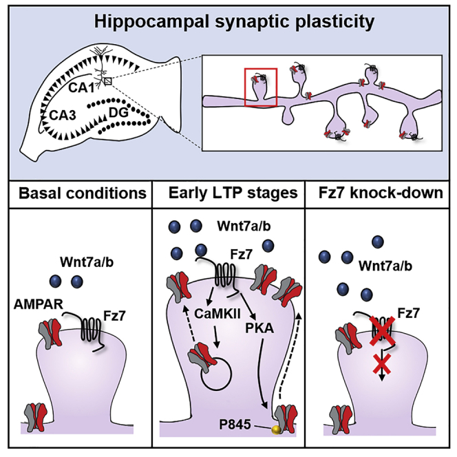
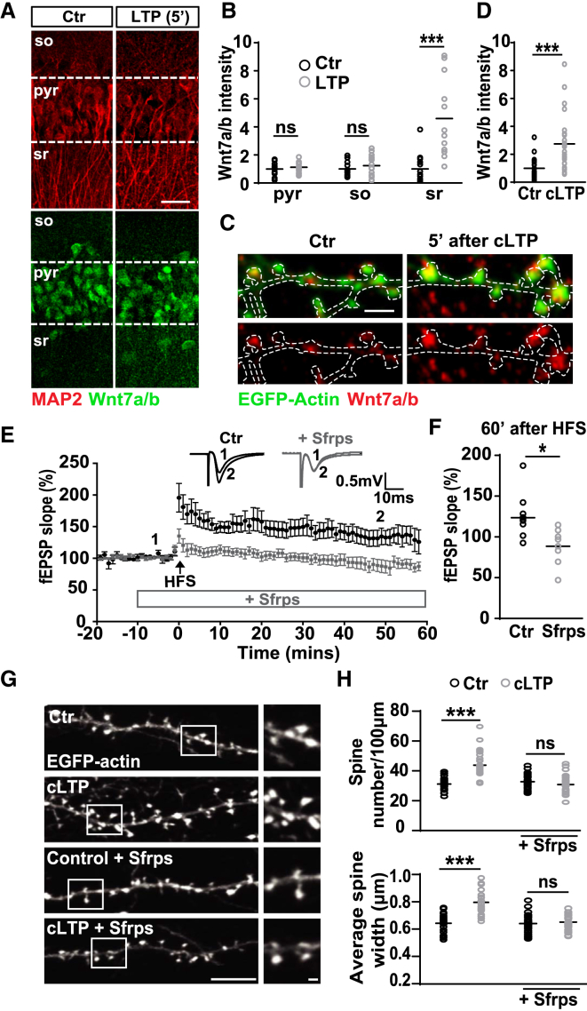
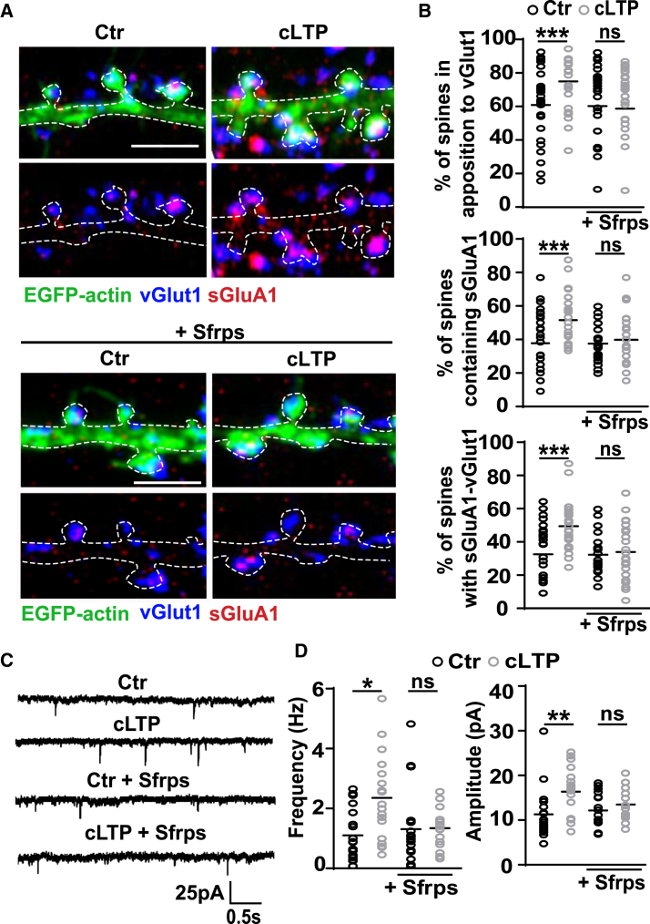
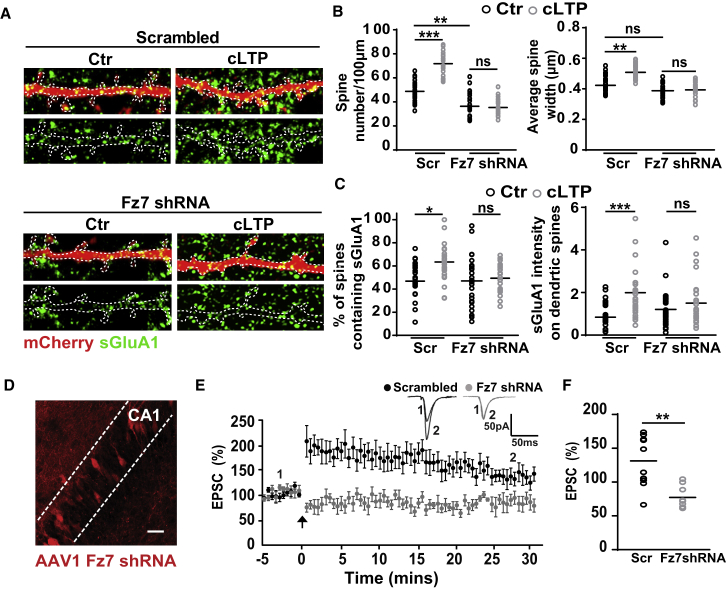
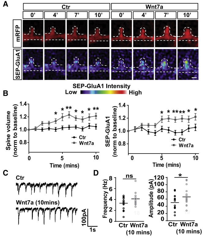
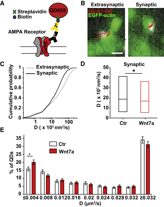
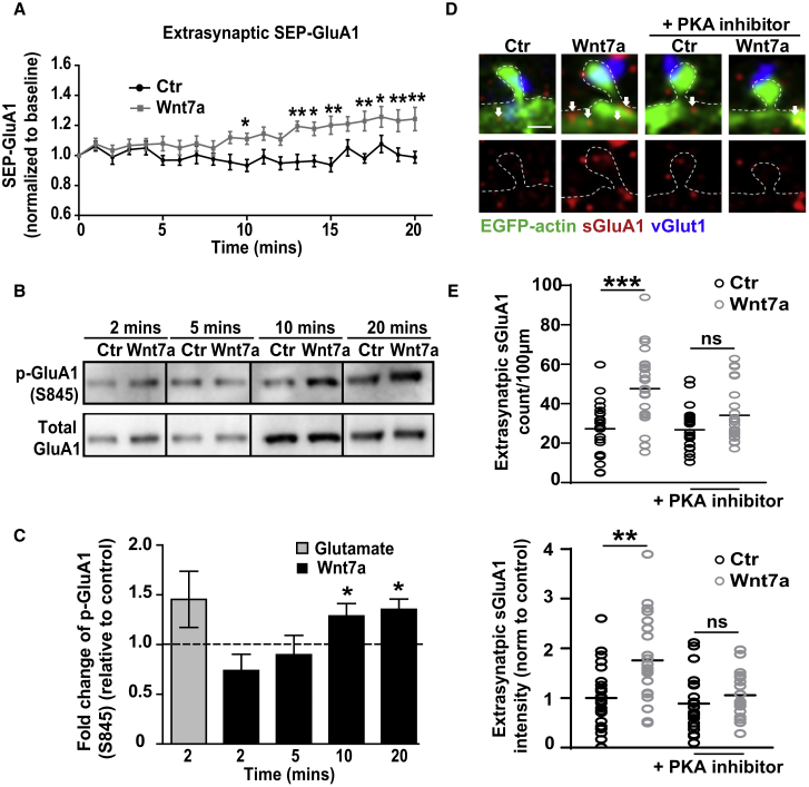
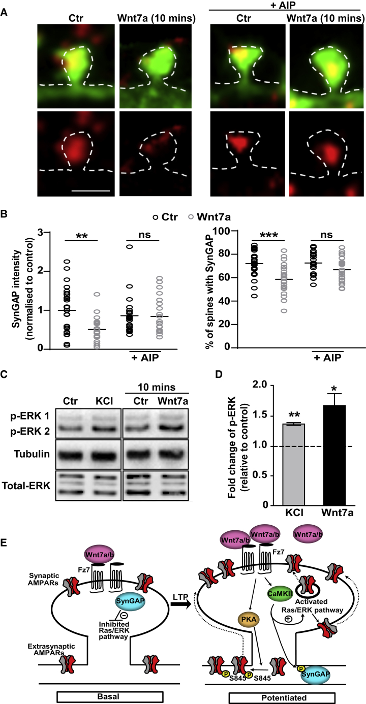
The structural and functional plasticity of synapses is critical for learning and memory. Long-term potentiation (LTP) induction promotes spine growth and AMPAR accumulation at excitatory synapses, leading to increased synaptic strength. Glutamate initiates these processes, but the contribution from extracellular modulators is not fully established. Wnts are required for spine formation; however, their impact on activity-mediated spine plasticity and AMPAR localization is unknown. We found that LTP induction rapidly increased synaptic Wnt7a/b protein levels. Acute blockade of endogenous Wnts or loss of postsynaptic Frizzled-7 (Fz7) receptors impaired LTP-mediated synaptic strength, spine growth, and AMPAR localization at synapses. Live imaging of SEP-GluA1 and single-particle tracking revealed that Wnt7a rapidly promoted synaptic AMPAR recruitment and trapping. Wnt7a, through Fz7, induced CaMKII-dependent loss of SynGAP from spines and increased extrasynaptic AMPARs by PKA phosphorylation. We identify a critical role for Wnt-Fz7 signaling in LTP-mediated synaptic accumulation of AMPARs and spine plasticity.

Keywords: AMPA receptors; Frizzled-7; LTP; Sfrps; Wnt signaling; spine plasticity; synaptic plasticity.


Figures

None
Graphical abstract
Figure 1
Figure 1
Wnt Proteins Are Upregulated and Required for Structural and Functional Plasticity during LTP (A) Endogenous Wnt7a/b protein (green) in the CA1 pyramidal cell layer (pyr), stratum oriens (so), and stratum radiatum (sr) regions from acute hippocampal slices in control (Ctr) and 5 min after LTP induction. MAP2 (red) used as a reference marker (scale bar: 25 μm). (B) Wnt7a/b fluorescence intensity in control and LTP-induced slices normalized to control levels. n = 11–15 slices from 3 animals (∗∗∗p < 0.01, Student’s t test). (C) EGFP-actin-expressing cultured neurons (14 DIV) exposed to control or cLTP conditions. Endogenous Wnt7a/b protein in red (scale bar: 2.5 μm). (D) Wnt7a/b fluorescence intensity (normalized to control) in spines measured 5 min after cLTP treatment. n = 24 cells per condition (∗∗∗p < 0.001, Student’s t test). (E) Impact on LTP elicited by HFS in control (black) or Sfrp-treated acute hippocampal slices (gray). Insets show averaged fEPSP recordings before HFS (1) and 50–60 min after HFS (2). Data expressed as mean ± SEM. (F) fEPSPs 60 min after HFS (average of last 10 min of recording) in control and Sfrp-treated slices. n = 8–9 slices from 3–4 animals for each group (p < 0.05, Student’s t test). (G) Different EGFP-actin-expressing cultured neurons (14 DIV) exposed to control (Ctr) or cLTP conditions in the presence or absence of Sfrps (scale bars: 10 and 1 μm on zoomed image). (H) Spine number (above) and spine width (below). n = 35–40 cells per condition (∗∗∗p < 0.001, ANOVA). See also Figures S1 and S2.
Figure 2
Figure 2
Wnt Blockade Suppresses Activity-Mediated Synaptic Localization of AMPARs (A) Cultured neurons (14 DIV) exposed to control (Ctr) or cLTP conditions, with or without Sfrps. Excitatory presynaptic marker vGlut1 (blue), surface excitatory postsynaptic marker sGluA1 (red), and EGFP-actin (green) (scale bars: 2.5 μm). (B) Percentage of spines containing sGluA1 and in apposition to vGlut1. Percentage of synapses was determined by the colocalization of vGlut1 and sGluA1. n = 39–52 cells per condition (∗∗∗p < 0.001, ANOVA). (C) AMPAR-mediated mEPSCs recorded from hippocampal neurons (14 DIV) exposed to control (Ctr) or cLTP conditions in the presence or absence of Sfrps. (D) mEPSC frequency and amplitude. n = 15–19 cells recorded in each group from 6 independent experiments (p < 0.05 and ∗∗p < 0.01, ANOVA). See also Figure S2.
Figure 3
Figure 3
Fz7 Receptors Are Required for Structural and Functional Plasticity and AMPAR Localization during LTP (A) Scrambled or Fz7 shRNA-transfected cultured neurons (14–16 DIV) exposed to control (Ctr) or cLTP conditions. sGluA1 (green) with mCherry-labeled spines (scale bars: 2.5 μm). (B) Spine number and width in neurons exposed to different conditions. n = 25–27 cells per condition (∗∗p < 0.01 and ∗∗∗p < 0.001, ANOVA). (C) Quantification of surface GluA1 levels at dendritic spines. n = 25–27 cells per condition (p < 0.05 and ∗∗∗p < 0.001, ANOVA). (D) Example image of an organotypic hippocampal slice infected with AAV1 driving the expression of Fz7 shRNA in the CA1 region (scale bar: 25 μm). (E) Sample traces (insets) and summary LTP graph from whole-cell patched neurons in organotypic slices infected with AAV-expressing scrambled or Fz7 shRNA following a pairing protocol (3 Hz stimulation during depolarization to 0 mV; arrow). Data expressed as mean ± SEM. (F) Percentage of EPSC response 30 min after the pairing protocol (average of last 10 min of recording) in scrambled (Scr) and Fz7 shRNA-infected organotypic slices. n = 8–10 cultured slices for each group from four independent cultures (∗∗∗p < 0.001, Student’s t test). See also Figures S3–S5 and S7.
Figure 4
Figure 4
Wnt7a Rapidly Increases AMPAR Localization on Spines and AMPAR Currents (A) mRFP and SEP-GluA1 (pseudo-color) signals in a spine (14 DIV) exposed to control (Ctr) or Wnt7a and imaged over time (scale bar: 1 μm). (B) mRFP volume and SEP-GluA1 intensity at different time points, normalized to baseline. n = 27–33 spines from 7–8 cells (p < 0.05 and ∗∗p < 0.01, ANOVA with repeated measures). Data expressed as mean ± SEM. (C) Spontaneous EPSCs from hippocampal neurons (13–14 DIV) treated with or without Wnt7a for 10 min. (D) Frequency and amplitude of spontaneous EPSC events. n = 18 cells recorded in each group from 5 independent experiments (p < 0.05, Student’s t test for EPSC amplitude). See also Figure S6.
Figure 5
Figure 5
Wnt7a Promotes the Diffusional Trapping of AMPARs in Dendritic Spines (A) Diagram of quantum dot-tagged GluA1 (QD-GluA1). Streptavidin-coupled quantum dots (QDs) recognize biotinylated secondary antibodies bound to GluA1-targeted primary antibodies. (B) Extrasynaptic (left panel) and synaptic (right panel) QD-GluA1 trajectories along the dendrites of hippocampal neurons (13–14 DIV). Spines are visualized with EGFP-actin (scale bar: 1 μm). (C) Cumulative probability of diffusion coefficient (D coefficient) in a log scale between synaptic and extrasynaptic QD-GluA1 in control conditions. (D) Median D coefficient at the synapse in control (Ctr) and Wnt7a-treated cells (10 min) (p < 0.05, Kolmogorov-Smirnov test). (E) Overall distribution of the D coefficient data shows significant change in the immobile fraction between control (Ctr) and Wnt7a (p < 0.05, Student’s t test. Data expressed as mean ± SEM. Entire dataset n = 1,638 (control) and 1,409 (Wnt7a) of synaptic trajectories and 12,350 (control) and 10,459 (Wnt7a) of extrasynaptic trajectories from 4 independent experiments. See also Figure S6.
Figure 6
Figure 6
Wnt7a Signals through PKA to Promote Extrasynaptic Localization of AMPARs (A) Extrasynaptic SEP-GluA1 intensity over time in neurons treated with control (Ctr) or Wnt7a, normalized to baseline values. n = 17 dendritic ROIs from 5 cells (p < 0.05 and ∗∗p < 0.01, ANOVA with repeated measures). (B) Phosphorylation of GluA1 at S845 (p-GluA1) following 2–20 min of Wnt7a treatment (14 DIV). (C) p-GluA1 levels normalized to total GluA1 levels. Graphs show fold changes in S845 levels relative to controls (dashed line) over time. Glutamate (50 μM) was used as a positive control. n = 4 experiments per dataset (p < 0.05, Student’s t test). Data expressed as mean ± SEM. (D) EGFP-actin-expressing hippocampal neurons (14 DIV) exposed to control (Ctr) or Wnt7a for 20 min with or without the PKA inhibitor PKI (14–22) amide. vGlut1 (blue) and sGluA1 (red); arrows indicate extrasynaptic sGluA1 puncta (scale bar: 1 μm). (E) Extrasynaptic sGluA1 puncta normalized to dendritic length, and intensity of extrasynaptic sGluA1 normalized to control. n = 23–27 cells per condition (∗∗p < 0.01 and ∗∗∗p < 0.001, ANOVA). See also Figure S7.
Figure 7
Figure 7
Wnt7a-Mediated Reduction of SynGAP in Spines and Activation of the Ras-ERK Pathway (A) Spines from EGFP-actin-expressing neurons (14 DIV) with endogenous SynGAP (red) exposed to control (Ctr) or Wnt7a for 10 min with or without the CaMKII inhibitor AIP (scale bar: 1 μm). (B) SynGAP intensity within small to medium spines normalized to control levels, and the percentage of spines containing SynGAP. n = 25 cells per condition (∗∗p < 0.01 and ∗∗∗p < 0.001, ANOVA). (C) Phosphorylated ERK (p-ERK1 and p-ERK2) 10 min after exposure to Wnt7a (14 DIV). Tubulin was used as a loading control. (D) Quantification of p-ERK levels normalized to total ERK levels. Graphs show fold change in p-ERK levels relative to controls (dashed line). Potassium chloride (KCl; 60 mM) was used as a positive control (p < 0.05 and ∗∗p < 0.01, Student’s t test). Data expressed as mean ± SEM. (E) Proposed model for the mechanism by which Wnt7a-Fz7 signaling regulates LTP-induced AMPAR localization and spine growth. LTP induction increases endogenous Wnt7a/b levels at synapses. Wnt7a/b binding to Fz7 activates PKA and CaMKII, resulting in increased levels of extrasynaptic and synaptic AMPARs through the loss of SynGAP at synapses. Dashed arrows represent potential mechanisms. See also Figures S6 and S7.

Similar articles

Cited by

References

    1. Araki Y., Zeng M., Zhang M., Huganir R.L. Rapid dispersion of SynGAP from synaptic spines triggers AMPA receptor insertion and spine enlargement during LTP. Neuron. 2015;85:173–189. - PMC - PubMed
    1. Ataman B., Ashley J., Gorczyca M., Ramachandran P., Fouquet W., Sigrist S.J., Budnik V. Rapid activity-dependent modifications in synaptic structure and function require bidirectional Wnt signaling. Neuron. 2008;57:705–718. - PMC - PubMed
    1. Barria A., Malinow R. NMDA receptor subunit composition controls synaptic plasticity by regulating binding to CaMKII. Neuron. 2005;48:289–301. - PubMed
    1. Bassani S., Folci A., Zapata J., Passafaro M. AMPAR trafficking in synapse maturation and plasticity. Cell. Mol. Life Sci. 2013;70:4411–4430. - PMC - PubMed
    1. Borgdorff A.J., Choquet D. Regulation of AMPA receptor lateral movements. Nature. 2002;417:649–653. - PubMed

Publication types

MeSH terms