. 2018 Apr 24;23(4):1152-1165.
doi: 10.1016/j.celrep.2018.03.043.

Cell Autonomous and Non-cell Autonomous Regulation of SMC Progenitors in Pulmonary Hypertension

Affiliations

Cell Autonomous and Non-cell Autonomous Regulation of SMC Progenitors in Pulmonary Hypertension

Abdul Q Sheikh et al. Cell Rep. .

Abstract

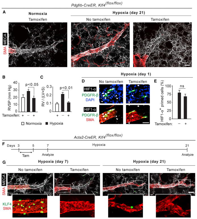
Pulmonary hypertension is a devastating disease characterized by excessive vascular muscularization. We previously demonstrated primed platelet-derived growth factor receptor β+ (PDGFR-β+)/smooth muscle cell (SMC) marker+ progenitors at the muscular-unmuscular arteriole border in the normal lung, and in hypoxia-induced pulmonary hypertension, a single primed cell migrates distally and expands clonally, giving rise to most of the pathological smooth muscle coating of small arterioles. Little is known regarding the molecular mechanisms underlying this process. Herein, we show that primed cell expression of Kruppel-like factor 4 and hypoxia-inducible factor 1-α (HIF1-α) are required, respectively, for distal migration and smooth muscle expansion in a sequential manner. In addition, the HIF1-α/PDGF-B axis in endothelial cells non-cell autonomously regulates primed cell induction, proliferation, and differentiation. Finally, myeloid cells transdifferentiate into or fuse with distal arteriole SMCs during hypoxia, and Pdgfb deletion in myeloid cells attenuates pathological muscularization. Thus, primed cell autonomous and non-cell autonomous pathways are attractive therapeutic targets for pulmonary hypertension.

Keywords: cardiovascular disease; endothelial-smooth muscle cell interactions; pulmonary artery; pulmonary hypertension; pulmonary vascular disease; smooth muscle biology; smooth muscle progenitors; vascular biology; vascular wall; vasculoproliferative disease.


Conflict of interest statement

DECLARATION OF INTERESTS

The authors declare no competing interests.

Figures

Figure 1
Figure 1. HIF1-α Is Required for Distal Arteriole Muscularization and PH
(A) Wild-type mice were exposed to normoxia or hypoxia (10% FiO2) for 1 day, and then lung vibratome sections were stained for PDGFR-β, SMA, HIF1-α, and DAPI as indicated. The muscular-unmuscular borders of pulmonary arterioles near airway branch L.L1.A1 (left bronchus-first lateral secondary branch-first anterior branches) are shown. The boxed regions in hypoxia are shown as close-ups on the right, with arrowheads indicating HIF1-α + primed cells. (B) Percentage of primed cells expressing HIF1-α at hypoxia day 1 is shown. n = 5 lungs, with 2–3 arterioles per lung. Total primed cells scored were 39 in normoxia and 37 in hypoxia. (C–G) Pdgfrb-CreERT2, Hif1a(flox/flox) mice were injected with tamoxifen (1 mg/day for 5 days), rested, and then exposed to normoxia or hypoxia for 21 days (C–E) or 2 days (F and G). Arterioles were stained for SMA and panendothelial cell antigen antibody (MECA-32) in (C) and for PDGFR-β, KLF4, SMA, and DAPI in (F), as indicated. Right ventricle systolic pressure (RVSP) and the ratio of the weight of the right ventricle (RV) to that of the sum of the left ventricle (LV) and septum (S) are shown (D and E); n = 4 mice. The percentage of primed cells that are KLF4+ at hypoxia day 2 is quantified in (G); n = 4 mice. Total primed cells scored were 30 and 32 in no-tamoxifen and tamoxifen groups, respectively. NS, not significant. (H and I) Arterioles of Pdgfrb-CreERT2, Hif1a (flox/flox), ROSA26R (mTmG/+) mice were stained for GFP (lineage tag), SMA, and CD68 after 14 days of hypoxia in (H). The percentage of distal SMA+ cells expressing GFP or CD68 are quantified in (I). n = 3 mice and 3 arterioles per lung. 35 SMA+ cells were scored. (J) Experimental strategy for (K), in which Pdgfrb-CreERT2, Hif1a(flox/flox) mice are induced with tamoxifen (1.5 mg/day) on days 3–5 of hypoxia. (K) Arterioles were stained with SMA and MECA-32. n = 5 mice and 2–3 arterioles per lung. All error bars indicate SD. Scale bars, 20 μm. See also Figures S1–S3.
Figure 2
Figure 2. Primed Cell KLF4 Is Required for Distal Muscularization and PH
(A–E) Pdgfrb-CreERT2, Klf4(flox/flox) mice were induced with tamoxifen (1 mg/day for 5 days), rested for 5 days, and then exposed to normoxia or hypoxia for 1 day (D and E) or 21 days (A–C). Arterioles near L.L1.A1 were stained for SMA and MECA-32 in (A) and for HIF1-α, PDGFR-β, and DAPI in (D). RVSP and RV weight ratio are shown in (B and C), respectively; n = 5 mice. The percentage of HIF1-α + primed cells at hypoxia day 1 is shown in (E); n = 4 mice, with 2–3 arterioles per lung. Total primed cells scored were 26 and 29 in no-tamoxifen and tamoxifen groups, respectively. ns, not significant. (F) Experimental strategy for (G), in which Acta2-CreERT2, Klf4(flox/flox) mice are induced with tamoxifen (1.5 mg/day) from hypoxia days 3–5. (G) Arterioles of Acta2-CreERT2; Klf4(flox/flox) mice were stained with SMA, MECA-32, and KLF4 after days 7 or 21 of hypoxia. n = 5 mice, with 2–3 arteriole per lung. All error bars indicate SD. Scale bars, 20 μm. See also Figure S2.
Figure 3
Figure 3. Endothelial Hif1a Deletion Attenuates Distal Muscularization and PH and Perturbs SMC Differentiation
Cdh5-CreERT2, Hif1a(flox/flox) mice were injected with tamoxifen (1 mg/day for 5 days), rested for 5 days, and then exposed to normoxia or hypoxia for indicated duration. Arterioles near L.L1.A1 airway branches were analyzed in lung vibratome sections stained for SMA. Sections were also stained for PDGF-B and MECA-32 in (A); PDGFR-β, KLF4, and DAPI in (D); PDGFR-β and HIF1-α in (F); and SMMHC, PDGFR-β, and DAPI in (H). (B and C) RVSP and weight ratio are shown; n = 4 mice. The percentage of primed cells at hypoxia day 2 that are KLF4+ is quantified in (E); n = 4 mice. Total primed cells scored were 33 and 30 in no-tamoxifen and tamoxifen groups, respectively. In (G), the percentage of SMA+PDGFR-β + cells at hypoxia day 1 (primed cells) or hypoxia day 5 (middle or distal arteriole SMCs) that are also HIF1-α + is shown. n = 4 mice. 31 cells were scored at hypoxia day 1 for the no-tamoxifen group and 32 cells were scored for the tamoxifen group, whereas 152 cells were scored at hypoxia day 5 for the no-tamoxifen group and 160 cells were scored for the tamoxifen group. The percentage of distal arteriole SMA+ cells that are PDGFR-β + and/or SMMHC+ at hypoxia day 7 is shown in (I). n = 5 mice. 235 cells with and 219 cells without tamoxifen were scored for PDGFR-β, and 72 cells with and 53 cells without tamoxifen were scored for SMMHC. *p < 0.05 versus tamoxifen, ^p < 0.05 versus hypoxia day 1, ~p < 0.01 versus SMMHC+ cells. ns, not significant. All error bars indicate SD. Scale bars, 20 μm. See also Figures S2 and S4.
Figure 4
Figure 4. Endothelial-Specific Vhl Deletion Induces HIF1-α and PDGF-B Expression and Distal Arteriole Muscularization under Normoxia
Cdh5-CreERT2, Vhl(flox/flox) mice were injected with tamoxifen (1 mg/day for 5 days) and then analyzed for 12 days (A and B) or 47 days (C and D) thereafter. (A–C) Arterioles near L.L1.A1 airways were analyzed in vibratome sections stained for SMA, as well as for HIF1-α and MECA-32 in (A), PDGF-B and KLF4 in (B), or PDGFR-β, KLF4, and nuclei (DAPI) in (C). Boxed regions are shown as close-ups. n = 4 mice, with 2–3 arterioles per mouse. (D) Quantification of the percentage of KLF4+ primed cells 1 week after tamoxifen induction from images shown in (C). n = 5 mice, with 2–3 arterioles per mouse. Total primed cells scored were 45 and 41 in no-tamoxifen and tamoxifen groups, respectively. All error bars indicate SD. Scale bars, 20 μm. See also Figure S2.
Figure 5
Figure 5. Endothelial-Specific Pdgfb Deletion Attenuates Distal Muscularization, KLF4 Expression, and PH and Perturbs SMC Differentiation
(A) Wild-type and Pdgfb(+/−) mice were exposed to normoxia or hypoxia for 1 day, and then lung vibratome sections were stained for SMA, HIF1-α, PDGFR-β, and DAPI. (B) Quantification of primed cells that express HIF1-α at day 1 of hypoxia. n = 5 mice of each genotype, with 2–3 arterioles per mouse. 42 and 39 primed cells were scored in wild-type and Pdgfb(+/−) lungs, respectively. (C–I) Cdh5-CreERT2, Pdgfb(flox/flox) mice were injected with tamoxifen (1 mg/day for 5 days), rested for 5 days, and then exposed to normoxia or hypoxia for 2, 7, or 21 days as indicated. Distal arterioles near L.L1.A1 airway branches were stained with SMA, as well as for MECA-32 in (C), KLF4 and PDGFR-β in (F), or SMMHC and PDGFR-β in (H). RVSP and RV weight ratio are quantified in (D) and (E), respectively. n = 5 mice. In (G), the percentage of KLF4+ primed cells at hypoxia day 2 was quantified. n = 5 mice, with 2–3 arteriole per lung. Total primed cells scored were 37 and 41 in no-tamoxifen and tamoxifen groups, respectively. In (I), the percentage of SMA+ distal arteriole cells at hypoxia day 7 that are PDGFR-β + and/or SMMHC+ are quantified. n = 5 mice. In the distal arteriole, 233 cells with and 217 cells without tamoxifen were scored for PDGFR-β, and 78 cells with and 71 cells without tamoxifen were scored for SMMHC. *p < 0.05 versus tamoxifen treatment, ~p < 0.01 versus SMMHC+ cells. All error bars indicate SD. Scale bars, 20 μm. See also Figures S2, S4, and S5.
Figure 6
Figure 6. Macrophages Give Rise to Distal Arteriole SMC Marker+ Cells, and Macrophage-Derived PDGF-B Is Integral for Distal Muscularization
Tamoxifen (1 mg/day) was injected for 5 days in Acta2-CreERT2 (C) or Cdh5-CreERT2 (D and E) mice or for 15 days in Csf1r-Mer-iCre-Mer mice (A, B, and F). Mice were rested and exposed to normoxia or hypoxia for 7, 19, or 21 days as indicated, and then vibratome sections containing distal arterioles near L.L1.A1 airway branches were immunostained. (A–C) Sections from ROSA26R(mTmG/+) mice carrying Csf1r-Mer-iCre-Mer (A and B) or Acta2-CreERT2 (C) were stained for GFP (lineage tag), CD68, nuclei (DAPI), and either SMA (A and C) or SMMHC (B). Boxed regions are shown as close-ups. In (A), arrowhead points to a SMA+ cell expressing GFP and CD68, and the asterisk indicates the SMA+GFP+CD68 cell. Close-ups in (B) show SMMHC+GFP+CD68 cells and in (C) show SMA cells that are GFPCD68+. (D and E) Sections from Cdh5-CreERT2 mice that are homozygous for floxed Hif1a (D) or Pdgfb (E) alleles were stained for PDGF-B, SMA, and CD68. (F) Sections from Csf1r-Mer-iCre-Mer, Pdgfb(flox/flox) mice were stained for PDGF-B, SMA, and MECA-32. Boxed regions are shown as close-ups below. n = 3 mice, with 2–3 arterioles per mouse. Scale bars, 20 μm. See also Figures S2 and S6.
Figure 7
Figure 7. Summary of Cell Autonomous and Non-cell Autonomous Regulation of Primed Cells in Hypoxia-Induced Distal Pulmonary Arteriole Muscularization
During hypoxia days 1–3, EC expression of HIF1-α and PDGF-B induces primed cell upregulation of KLF4 (progenitor induction). This progenitor induction results in initial migration of a primed cell beyond the M (middle)-to-D (distal) arteriole border. In contrast to EC HIF1-α, primed cell HIF1-α is expendable for progenitor induction and initial migration. Distal arteriole SMC proliferation peaks at hypoxia day 7 (Sheikh et al., 2014), and at hypoxia days 5–7, lung PDGF-B expression by ECs, and perhaps by macrophages, stays at high levels, resulting in HIF1-α expression in distal arteriole SMCs. Expansion of the primed cell lineage in the distal arteriole requires this lineage to express HIF1-α, but not KLF4. During hypoxia days 14–21, PDGF-B expression in the lung wanes and newly derived distal arteriole cells differentiate into mature SMCs. Furthermore, pre-existing macrophages transdifferentiate into or fuse with distal arteriole SMCs.

References

    1. Amsellem V, Abid S, Poupel L, Parpaleix A, Rodero M, Gary-Bobo G, Latiri M, Dubois-Rande JL, Lipskaia L, Combadiere C, Adnot S. Roles for the CX3CL1/CX3CR1 and CCL2/CCR2 chemokine systems in hypoxic pulmonary hypertension. Am J Respir Cell Mol Biol. 2017;56:597–608. - PubMed
    1. Angelini DJ, Su Q, Kolosova IA, Fan C, Skinner JT, Yamaji-Kegan K, Collector M, Sharkis SJ, Johns RA. Hypoxia-induced mitogenic factor (HIMF/FIZZ1/RELM alpha) recruits bone marrow-derived cells to the murine pulmonary vasculature. PLoS ONE. 2010;5:e11251. - PMC - PubMed
    1. Arias-Stella J, Saldana M. The terminal portion of the pulmonary arterial tree in people native to high altitudes. Circulation. 1963;28:915–925. - PubMed
    1. Ball MK, Waypa GB, Mungai PT, Nielsen JM, Czech L, Dudley VJ, Beussink L, Dettman RW, Berkelhamer SK, Steinhorn RH, et al. Regulation of hypoxia-induced pulmonary hypertension by vascular smooth muscle hypoxia-inducible factor-1α. Am J Respir Crit Care Med. 2014;189:314–324. - PMC - PubMed
    1. Brusselmans K, Compernolle V, Tjwa M, Wiesener MS, Maxwell PH, Collen D, Carmeliet P. Heterozygous deficiency of hypoxia-inducible factor-2alpha protects mice against pulmonary hypertension and right ventricular dysfunction during prolonged hypoxia. J Clin Invest. 2003;111:1519–1527. - PMC - PubMed

Publication types

MeSH terms