. 2018 Apr 24;23(4):974-982.
doi: 10.1016/j.celrep.2018.03.104.

Obesity Suppresses Cell-Competition-Mediated Apical Elimination of RasV12-Transformed Cells from Epithelial Tissues

Affiliations

Obesity Suppresses Cell-Competition-Mediated Apical Elimination of RasV12-Transformed Cells from Epithelial Tissues

Ayana Sasaki et al. Cell Rep. .

Abstract

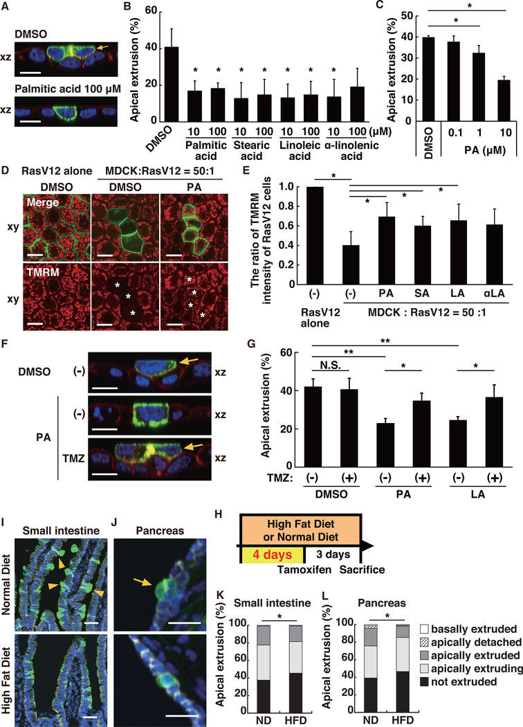
Recent studies have revealed that newly emerging transformed cells are often eliminated from epithelial tissues via cell competition with the surrounding normal epithelial cells. This cancer preventive phenomenon is termed epithelial defense against cancer (EDAC). However, it remains largely unknown whether and how EDAC is diminished during carcinogenesis. In this study, using a cell competition mouse model, we show that high-fat diet (HFD) feeding substantially attenuates the frequency of apical elimination of RasV12-transformed cells from intestinal and pancreatic epithelia. This process involves both lipid metabolism and chronic inflammation. Furthermore, aspirin treatment significantly facilitates eradication of transformed cells from the epithelial tissues in HFD-fed mice. Thus, our work demonstrates that obesity can profoundly influence competitive interaction between normal and transformed cells, providing insights into cell competition and cancer preventive medicine.

Keywords: Aspirin; EDAC; RasV12; apical extrusion; cancer prevention; cell competition; chronic inflammation; lipid metabolism; obesity; pancrease.


Conflict of interest statement

DECLARATION OF INTERESTS

The authors declare no competing interests.

Figures

Figure 1.
Figure 1.. HFD Treatment Suppresses Apical Elimination of RasV12-Transformed Cells from the Epithelial Monolayer of the Small Intestine and Pancreas, but Not of the Lung
(A) Strategy for the establishment of the cell competition mouse model. (B) Experimental design for feeding and tamoxifen administration. (C, E, and G) Immunofluorescence images of RasV12-transformed cells in the epithelium of the small intestine (C), pancreas (E), and lung (G). The tissue samples were stained with anti-GFP (green) and anti-E-cadherin (gray) antibodies and Hoechst (blue). The yellow arrow and arrowheads indicate apically extruded and extruding cells, respectively. The scale bars represent 50 μm (left panels) and 20 μm (right panels). (D, F, and H) Quantification of apical extrusion of RasV12 cells for C, E, and G, respectively. (D) ND 2,063 cells from 8 mice; HFD 1,117 cells from 3 mice. (F) ND 560 cells from 9 mice; HFD 298 cells from 4 mice. (H) ND 144 cells from 4 mice; HFD 213 cells from 4 mice. *p < 0.0001 (chi-square test). N.S., not significant.
Figure 2.
Figure 2.. Alteration of Lipid Metabolism Affects Apical Extrusion of RasV12-Transformed Cells
(A and B) Effect of various fatty acids on apical extrusion of RasV12-transformed cells. MDCK-pTR GFP-RasV12 cells were mixed with normal MDCK cells on collagen gels. Cells were cultured with the indicated concentration of fatty acids and fixed after 24 hr incubation with tetracycline and stained with Alexa-Fluor-568-phalloidin (red) and Hoechst (blue) (C) Dose-dependent effect of palmitic acid on apical extrusion of RasV12 cells. (D and E) Effect of various fatty acids on TMRM incorporation in RasV12-transformed cells surrounded by normal cells. MDCK-pTR GFP-RasV12 cells were mixed with normal MDCK cells on collagen gels. Cells were cultured with the indicated fatty acid (100 μM), and TMRM incorporation was examined after 16 hr incubation with tetracycline. (F and G) Effect of the fatty acid oxidation inhibitor trimetazidine (TMZ) on apical extrusion of RasV12 cells. TMZ was added together with tetracycline and the fatty acid (100 μM) where indicated. (A, D, and F) Confocal microscopy images of xz (A and F) and xy (D) sections. Arrows indicate the apically extruded cells. Asterisks indicate RasV12 cells surrounded by normal cells. The scale bars represent 10 μm. (B, C, E, and G) Quantification of apical extrusion (B, C, and G) and the fluorescence intensity of TMRM (E). n ≥ 100 cells (B, C, and G) or n ≥ 10 cells (E) for each experimental condition is shown. Data are mean ± SD from three independent experiments. *p < 0.05; **p < 0.01 (Student’s t tests). (H–L) Effect of short-term HFD feeding on apical extrusion in vivo. (H) Experimental design for short-term HFD feeding and tamoxifen administration. (I and J) Immunofluorescence images of RasV12-transformed cells in the epithelium of the small intestine (I) and pancreas (J). The tissue samples were stained with anti-GFP (green) and anti-E-cadherin (gray) antibodies and Hoechst (blue). The arrow and arrowheads indicate apically extruded and extruding cells, respectively. The scale bars represent 50 μm (I) and 20 μm (J). (K and L) Quantification of apical extrusion of RasV12 cells in the small intestine (K) and pancreas (L). ND 940 cells from 3 mice; HFD 749 cells from 4 mice. ND 222 cells from 3 mice; HFD 348 cells from 4 mice. *p < 0.05 (chi-square test).
Figure 3.
Figure 3.. Chronic Inflammation Is Involved in HFD-Mediated Suppression of Apical Extrusion of RasV12-Transformed Cells
(A–E) Effect of the soybean-oil- or linseed-oil-enriched diet on apical extrusion in vivo. (A) Experimental design for diet feeding and tamoxifen administration. (B and D) Immunofluorescence images of RasV12-transformed cells in the epithelium of the small intestine (B) and pancreas (D). The tissue samples were stained with anti-GFP (green) and anti-E-cadherin (gray) antibodies and Hoechst (blue). Arrowheads indicate apically extruding cells. The scale bars represent 50 μm (B) and 20 μm (D). (C and E) Quantification of apical extrusion of RasV12 cells in the small intestine (C) and pancreas (E). (C) ND 2,863 cells from 8 mice, Soy 1,584 cells from 4 mice, and Lin 1,215 cells from 4 mice. (E) ND 560 cells from 9 mice, Soy 72 cells from 4 mice, and Lin 190 cells from 4 mice. *p < 0.05; **p < 0.0001 (chi-square test). (F–J) Effect of aspirin on apical extrusion of RasV12-transformed cells in HFD-fed mice. (F) Experimental design for diet feeding, aspirin treatment, and tamoxifen administration. (G and I) Immunofluorescence images of RasV12 cells in the epithelium of the small intestine (G) and pancreas (I). The arrow and arrowheads indicate apically extruded and extruding cells, respectively. The scale bars represent 30 μm (G) and 20 μm (I). (H and J) Quantification of apical extrusion of RasV12 cells in the small intestine (H) and pancreas (J). (H) ND (W, water) 481 cells, ND (Asp, aspirin) 517 cells, HFD (W) 600 cells, and HFD (Asp) 372 cells. (J) ND (W) 153 cells, ND (Asp) 237 cells, HFD (W) 212 cells, and HFD (Asp) 216 cells. For each condition, cells were collected from 3 mice. **p < 0.0001 (chi-square test).
Figure 4.
Figure 4.. In HFD-Fed Mice, RasV12-Transformed Cells Form Tumorous Lesions in the Pancreas
(A–C) The fate of RasV12-transformed cells after one month of tamoxifen administration in the pancreas in ND- or HFD-fed mice. (A) Experimental design for diet feeding and tamoxifen administration. (B) Immunofluorescence images of RasV12-transformed cells in the pancreas. The tissue samples were stained with anti-GFP (green) and anti-E-cadherin (gray) antibodies and Hoechst (blue). The dotted lines delineate the basement membrane of pancreatic epithelia. Arrows indicate basally extruded cells. The scale bars represent 50 μm. (C) Quantification of pancreatic epithelial ducts harboring RasV12-expressing cells. The percentage of ducts containing GFP-RasV12 cells relative to total ducts is shown. The total number of analyzed ducts are as follows: 198 (ND 3 days); 139 (HFD 3 days); 428 (ND 1 month); and 354 (HFD 1 month). Data are mean ± SD from three mice. *p < 0.05 (Student’s t tests).

References

    1. Afshin A, Forouzanfar MH, Reitsma MB, Sur P, Estep K, Lee A, Marczak L, Mokdad AH, Moradi-Lakeh M, Naghavi M, et al.; GBD 2015 Obesity Collaborators (2017). Health effects of overweight and obesity in 195 countries over 25 years. N. Engl. J. Med 377, 13–27. - PMC - PubMed
    1. Beyaz S, Mana MD, Roper J, Kedrin D, Saadatpour A, Hong SJ, Bauer-Rowe KE, Xifaras ME, Akkad A, Arias E, et al. (2016). High-fat diet enhances stemness and tumorigenicity of intestinal progenitors. Nature 531, 53–58. - PMC - PubMed
    1. Bhaskaran K, Douglas I, Forbes H, dos-Santos-Silva I, Leon DA, and Smeeth L (2014). Body-mass index and risk of 22 specific cancers: a population-based cohort study of 5$24 million UK adults. Lancet 384, 755–765. - PMC - PubMed
    1. Bianchini F, Kaaks R, and Vainio H (2002). Overweight, obesity, and cancer risk. Lancet Oncol 3, 565–574. - PubMed
    1. Bondar T, and Medzhitov R (2010). p53-mediated hematopoietic stem and progenitor cell competition. Cell Stem Cell 6, 309–322. - PMC - PubMed

Publication types

MeSH terms

LinkOut - more resources