Review
. 2018 Apr 24;7(5):88.
doi: 10.3390/jcm7050088.

Multi-Detector Computed Tomography Imaging Techniques in Arterial Injuries

Affiliations
Review

Multi-Detector Computed Tomography Imaging Techniques in Arterial Injuries

Cameron Adler et al. J Clin Med. .

Abstract

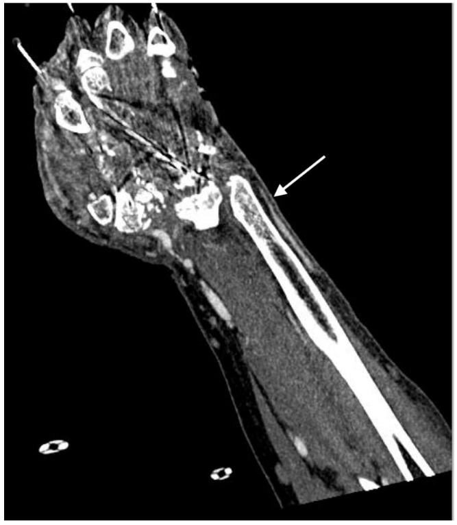
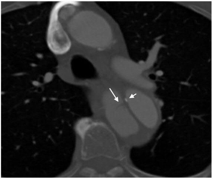
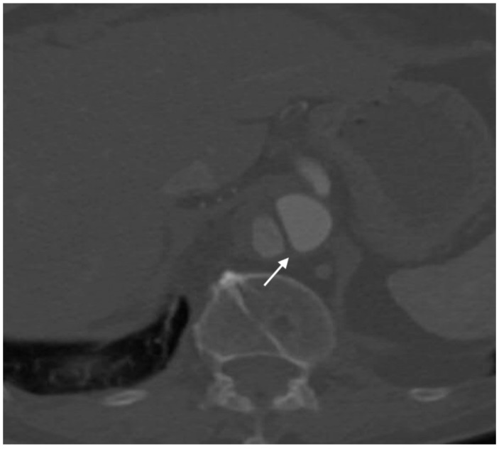
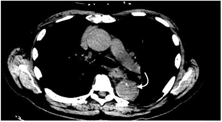
Cross-sectional imaging has become a critical aspect in the evaluation of arterial injuries. In particular, angiography using computed tomography (CT) is the imaging of choice. A variety of techniques and options are available when evaluating for arterial injuries. Techniques involve contrast bolus, various phases of contrast enhancement, multiplanar reconstruction, volume rendering, and maximum intensity projection. After the images are rendered, a variety of features may be seen that diagnose the injury. This article provides a general overview of the techniques, important findings, and pitfalls in cross sectional imaging of arterial imaging, particularly in relation to computed tomography. In addition, the future directions of computed tomography, including a few techniques in the process of development, is also discussed.

Keywords: CT; angiography; arterial injury; computed tomography; cross-sectional; imaging; radiology.


Conflict of interest statement

The authors declare no conflict of interest.

Figures

Figure 1
Figure 1
Consecutive axial images acquired during bolus tracking. The region of interest was placed over the ascending aorta, as marked by an asterisk. (a) Ascending aorta with attenuation greater than 50 Hounsfield units; (b) Ascending aorta with attenuation greater than 50 and less than 100 Hounsfield units; (c) Ascending aorta with attenuation greater than 120 Hounsfield units, at which point image acquisition began.
Figure 2
Figure 2
Volume rendering examples: (a) Volume rendering of distal radial artery occlusion; (b) Volume rendering of abdominal arteries.
Figure 3
Figure 3
Axial images of active extravasation of contrast into a medial left thigh hematoma following traumatic injury to the left leg: (a) Small volume of contrast extravasation seen during the arterial phase (arrow); (b) Increase in size of the collection of extravasated contrast in the delayed phase imaging (arrow).
Figure 4
Figure 4
Coronal image of right common femoral artery occlusion (arrow) with distal reconstitution (curved arrow) following traumatic injury to the pelvis.
Figure 5
Figure 5
Coronal image of right distal ulnar artery stenosis following traumatic injury to the wrist and hand.
Figure 6
Figure 6
Axial image of type A aortic dissection demonstrating both the cobweb sign (long arrow) and eccentric calcification in the dissection flap (short arrow) in the descending aorta. Note that the eccentric calcification abuts the true lumen.
Figure 7
Figure 7
Axial image of aortic dissection demonstrating the beak sign (arrow). Note the acute angle between the dissection flap and the outer wall.
Figure 8
Figure 8
Axial non-contrast image demonstrating intramural hematoma (curved arrow) in the descending thoracic aorta. Note that the crescent-shaped intramural hematoma has higher attenuation than the thin portions of normal arterial wall surrounding it.
Figure 9
Figure 9
Axial contrast-enhanced arterial phase image demonstrating a crescent-shaped intramural hematoma (curved arrow) in the descending aorta.
Figure 10
Figure 10
Axial image demonstrating large left peroneal artery pseudoaneurysm (arrow) following traumatic injury to the lower extremity.
Figure 11
Figure 11
Coronal image of the bilateral lower extremities in the arterial phase following left calf trauma. There is opacification of the left femoral vein (curved arrow) in the arterial phase, indicative of arteriovenous fistula. Note that the right femoral vein (arrow) is not opacified.

References

    1. Múnera F., Soto J.A., Palacio D., Velez S.M., Medina E. Diagnosis of arterial injuries caused by penetrating trauma to the neck: Comparison of helical CT angiography and conventional angiography. Radiology. 2000;216:356–362. doi: 10.1148/radiology.216.2.r00jl25356. - DOI - PubMed
    1. Lell M.M., Anders K., Uder M., Klotz E., Ditt H., Vega-Higuera F., Boskamp T., Bautz W.A., Tomandl B.F. New Techniques in CT Angiography. RadioGraphics. 2006;26:S45–S62. doi: 10.1148/rg.26si065508. - DOI - PubMed
    1. Yoon W., Kim J.K., Jeong Y.Y., Seo J.J., Park J.G., Kang H.K. Pelvic arterial hemorrhage in patients with pelvic fractures: Detection with contrast-enhanced CT. Radiographics. 2004;24:1591–1606. doi: 10.1148/rg.246045028. - DOI - PubMed
    1. Rieger M., Mallouhi A., Tauscher T., Lutz M., Jaschke W.R. Traumatic arterial injuries of the extremities: Initial evaluation with MDCT angiography. Am. J. Roentgenol. 2006;186:656–664. doi: 10.2214/AJR.04.0756. - DOI - PubMed
    1. Hamilton J.D., Manickam Kumaravel B., Michael B.L. Multidetector CT Evaluation of Active Extravasation in Blunt Abdominal and Pelvic Trauma Patients. Radiographics. 2008;28:1603–1616. doi: 10.1148/rg.286085522. - DOI - PubMed

LinkOut - more resources