. 2018 Jun 29;92(14):e00550-18.
doi: 10.1128/JVI.00550-18. Print 2018 Jul 15.

Lack of Transmission of Chronic Wasting Disease to Cynomolgus Macaques

Affiliations

Lack of Transmission of Chronic Wasting Disease to Cynomolgus Macaques

Brent Race et al. J Virol. .

Abstract

Chronic wasting disease (CWD) is a fatal prion disease that can infect deer, elk, and moose. CWD was first recognized in captive deer kept in wildlife facilities in Colorado from 1967 to 1979. CWD has now been detected in 25 U.S. states, 2 Canadian provinces, South Korea, Norway, and Finland. It is currently unknown if humans are susceptible to CWD infection. Understanding the health risk from consuming meat and/or products from CWD-infected cervids is a critical human health concern. Previous research using transgenic mouse models and in vitro conversion assays suggests that a significant species barrier exists between CWD and humans. To date, reported epidemiologic studies of humans consuming cervids in areas where CWD is endemic have found no evidence to confirm CWD transmission to humans. Previously, we reported data from ongoing cross-species CWD transmission studies using two species of nonhuman primates as models. Squirrel monkeys (SM) and cynomolgus macaques (CM) were inoculated by either the intracerebral or oral route with brain homogenates from CWD-infected deer and elk containing high levels of infectivity. SM were highly susceptible to CWD infection, while CM were not. In the present study, we present new data for seven CWD-inoculated CM euthanized 11 to 13 years after CWD inoculation and eight additional uninoculated control CM. New and archival CM tissues were screened for prion infection by using the ultrasensitive real-time quaking-induced conversion (RT-QuIC) assay, immunohistochemistry, and immunoblotting. In this study, there was no clinical, pathological, or biochemical evidence suggesting that CWD was transmitted from cervids to CM.IMPORTANCE Chronic wasting disease (CWD) is a fatal prion disease found in deer, elk, and moose. Since it was first discovered in the late 1960s, CWD has now spread to at least 25 U.S. states, 2 Canadian provinces, South Korea, Norway, and Finland. Eradication of CWD from areas of endemicity is very unlikely, and additional spread will occur. As the range and prevalence of CWD increase, so will the potential for human exposure to CWD prions. It is currently unknown if CWD poses a risk to human health. However, determining this risk is critical to preventing a scenario similar to that which occurred when mad cow disease was found to be transmissible to humans. In the present study, we used cynomolgus macaque monkeys as a surrogate model for CWD transmission to humans. After 13 years, no evidence for CWD transmission to macaques was detected clinically or by using highly sensitive prion disease-screening assays.

Keywords: CWD strains; RT-QuIC; barrier; chronic wasting disease; cross-species transmission; cynomolgus macaques; prion; squirrel monkeys.


Figures

FIG 1
FIG 1
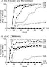
RT-QuIC analysis of Elk-1 (CWD inoculum) and vCJD brain from a clinically ill CM. RT-QuIC analysis using BV rPrPsen as a substrate was performed on serial 10-fold dilutions of CWD-infected elk brain (A) and vCJD-infected CM brain (B). Each curve represents the average fluorescence for four wells tested for each individual dilution. Dilutions are shown on the far right of each curve. In panel A, Elk-1 brain dilutions are indicated as ○ for a 10−4 dilution, ■ for 10−5, ▲ for 10−6, ▽ for 10−7, and □ for 10−8, and ● indicates normal deer brain diluted to 10−4 as a negative control. RT-QuIC using the BV rPrPsen substrate was highly sensitive for both CWD and cynomolgus macaque brain infected with vCJD. ND, not detected.
FIG 2
FIG 2
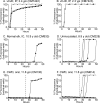
RT-QuIC analysis of vCJD-inoculated CM, CWD-inoculated CM, and uninoculated CM using the BV rPrPsen substrate. Four independent wells were tested for each CM, and each well is represented as an individual curve shown with one of four different symbols (○, ▲, ■, and ▽) in each panel. All tested brain samples were diluted to 10−3. (A and B) Positive-control vCJD-inoculated CM. (C) Normal elk brain-inoculated CM. (D) Uninoculated CM. (E and F) Two CWD-inoculated CM. The numbers of wells that reacted were variable between individual CM, but no consistent differences were observed between the uninoculated and CWD-inoculated groups. The vertical line shown at 25 h represents the cutoff time to meet the criteria for a positive reaction (see Results and Materials and Methods for more detail). IP, intraperitoneal; ypi, years postinfection; IC, intracerebral.
FIG 3
FIG 3
CM and SM brain histopathology. Shown are examples of gray matter regions from the cerebral cortex of an uninoculated CM (A, D, G, and J), CWD-inoculated CM (B, E, H, and K), and a CWD-infected squirrel monkey (C, F, I, and J). (A to C) Tissues were stained by H&E to look for spongiosis/vacuolation and general neuropathology. (D to L) Anti-GFAP IHC was used to detect activated astrocytes (D to F), and anti-PrP antibodies 6H4 and 3F4 were used with IHC to detect PrP deposition (G to L). No pathology, spongiform lesions, or excessive astroglial activation was observed in any of the uninoculated or CWD-inoculated CM brains. PrPsen could be seen in all brains as a smooth brown blush using anti-PrP antibody 6H4 (E and F). The background PrPsen was less noticeable using anti-PrP antibody 3F4 (G and H). The bar shown in panel A is 50 μm and applies to all panels.
FIG 4
FIG 4
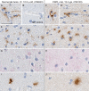
Immunohistochemical staining of CM brain using anti-PrP and -GFAP antibodies. Shown are examples of a normal elk brain-inoculated CM (A, B, E, G, and I) and a CWD-inoculated CM (C, D, F, H, and J). We tested three monoclonal anti-PrP antibodies (3F4, 6H4, and L42). All antibodies gave similar staining results, but only 3F4 is shown. Three patterns of aggregated PrP staining were observed: perivascular (A, C, and D), pericellular (E and F) and parenchymal (I and J). In all cases, aggregated PrP staining was rare and affected only small, focal regions of the brain. Panels G and H show minimal anti-GFAP staining and a lack of astrocytic activation in the same brain regions shown in panels E and F, respectively. A no-primary-antibody control (NP) is shown in panel B to demonstrate primary antibody staining specificity. The bar shown in panel A is 25 μm and applies to all panels.
FIG 5
FIG 5
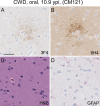
Immunohistochemical and H&E staining of the caudate nucleus from CM121. (A and B) Anti-PrP staining with the 3F4 antibody (A) and anti-PrP staining with the 6H4 antibody (B) show a cluster of PrP staining (brown). Several additional similar clusters were observed in the caudate nucleus of this monkey. (C) H&E staining of the same region depicted in panels A and B. No spongiform lesions were observed. (D) Minimal anti-GFAP staining and a lack of astrocytic activation in the same brain regions shown in panels A and B. The bar shown in panel A is 25 μm and applies to all panels.
FIG 6
FIG 6
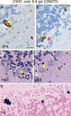
Hemosiderin pigment deposition in CM brain. All panels show a similar region of cerebellum from CWD-inoculated CM270. Staining methods are shown in the lower left corner of each panel. (A to D) Brown pigments (yellow arrows) were observed in slides stained with 3F4 (A), no-primary-antibody controls (B), and H&E-stained sections (C and D) of brain tissues examined from all the CWD-inoculated and uninoculated CM. (E) Prussian blue iron staining (blue) on adjacent sections confirmed that the pigment was hemosiderin. Hemosiderin pigment deposition was common near Purkinje cells, often near blood vessels (red arrow). The bar shown in panel A is 25 μm and applies to all panels.
FIG 7
FIG 7
Detection of PrP in spinal cords and nerve roots of CM using anti-PrP antibody (6H4). Spinal cord regions (cervical, thoracic, and lumbar) were screened for PrP deposition in 6 CWD-inoculated CM, 2 uninoculated CM, and 1 normal elk brain-inoculated CM. (A to E) Examples of thoracic spinal cord from one uninoculated CM (A), a normal elk brain-inoculated CM (C), and three CWD-inoculated CM (B, D, and E). PrP staining (brown) was similar for all CM. The strongest staining was located in the substantia gelatinosa region of the dorsal horn (blue arrow). (F and H) PrP staining (purple arrows) was also observed in nerve roots adjacent to the spinal cord. (G) The same nerve region depicted in panel F, without antibody 6H4 being applied (no primary ab). The bar shown in panel A is 1 mm and applies to panels A to E; the bar shown in panel F is 50 μm and applies to panels F to H.
FIG 8
FIG 8
Detection of astroglia in CM spinal cord using anti-GFAP antibody. Shown are an uninoculated CM (A, C, and D) and a CWD-inoculated CM (B, E, and F). (A and B) Typical astrocyte staining in spinal cord. (C to F) Higher magnifications show astrocyte staining in white matter (C and E) and gray matter (D and F), and squares drawn in panels A and B show the location of the magnified regions. No differences between CWD-inoculated and uninoculated CM were seen in astrocyte density, distribution, or cellular morphology. The bar in panel A is 1 mm and applies to panels A and B. The bar in panel C is 50 μm and applies to panels C to F.
FIG 9
FIG 9
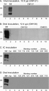
Immunoblot screening for PrPres in CWD-inoculated CM tissues. CWD-inoculated CM were euthanized at many different times throughout the 13.4-year study. Tissues from each individual CM were screened soon after euthanasia and again as a group at the conclusion of the study. All tissues shown were treated with proteinase K prior to testing. CWD-infected squirrel monkey brain (SM) was used as a positive control in each immunoblot. Lanes used for the molecular weight markers are marked with “mw.” Approximate molecular weights (in thousands) are shown at the left of each immunoblot. Immunoblots were probed with anti-PrP antibody 3F4 (A and B) and with anti-PrP antibody L42 (C and D). (A and B) Examples from two individual CM. (A) Lanes 3 to 10 show tissues from CM121 inoculated orally with CWD. Lanes: 3, cortex; 4, thalamus; 5, cerebellum; 6, spleen; 7, axillary lymph node; 8, cervical lymph node; 9, inguinal lymph node; 10, mesenteric lymph node. (B) Lanes 2 to 8 are tissues from CM130 inoculated orally with CWD, and lanes 9 to 10 are tissues from normal elk-inoculated CM633. Lanes: 2, ventral cerebral cortex; 3, hippocampus; 4, cerebellum; 5, brain stem; 6, dorsal cerebral cortex; 7, thalamus; 8, claustrum; 9, claustrum; 10, thalamus. (C) Cerebral cortex samples from 6 CM inoculated i.c. with CWD and 2 naive controls. (D) Cerebral cortex samples from 8 CM inoculated i.c. with CWD. Individual monkey identifications are provided across the top of panels C and D. In all blots, the SM positive-control lanes contained 0.24 mg and 0.36 mg of tissue equivalents, respectively. Experimental tissue lanes in panels A, C, and D contained 0.72 mg tissue equivalents per lane, and those in panel B contained 1.2 mg tissue equivalents per lane.

Similar articles

Cited by

References

    1. Saunders SE, Bartelt-Hunt SL, Bartz JC. 2012. Occurrence, transmission, and zoonotic potential of chronic wasting disease. Emerg Infect Dis 18:369–376. doi:10.3201/eid1803.110685. - DOI - PMC - PubMed
    1. Haley NJ, Hoover EA. 2015. Chronic wasting disease of cervids: current knowledge and future perspectives. Annu Rev Anim Biosci 3:305–325. doi:10.1146/annurev-animal-022114-111001. - DOI - PubMed
    1. Zabel M, Ortega A. 2017. The ecology of prions. Microbiol Mol Biol Rev 81:e00001-. doi:10.1128/MMBR.00001-17. - DOI - PMC - PubMed
    1. Uehlinger FD, Johnston AC, Bollinger TK, Waldner CL. 2016. Systematic review of management strategies to control chronic wasting disease in wild deer populations in North America. BMC Vet Res 12:173. doi:10.1186/s12917-016-0804-7. - DOI - PMC - PubMed
    1. Brown P, Cathala F, Raubertas RF, Gajdusek DC, Castaigne P. 1987. The epidemiology of Creutzfeldt-Jakob disease: conclusion of a 15-year investigation in France and review of the world literature. Neurology 37:895–904. doi:10.1212/WNL.37.6.895. - DOI - PubMed

Publication types

LinkOut - more resources