

Functional MRI in the Nile crocodile: a new avenue for evolutionary neurobiology
- PMID: 29695446
- PMCID: PMC5936729
- DOI: 10.1098/rspb.2018.0178
Functional MRI in the Nile crocodile: a new avenue for evolutionary neurobiology
Abstract
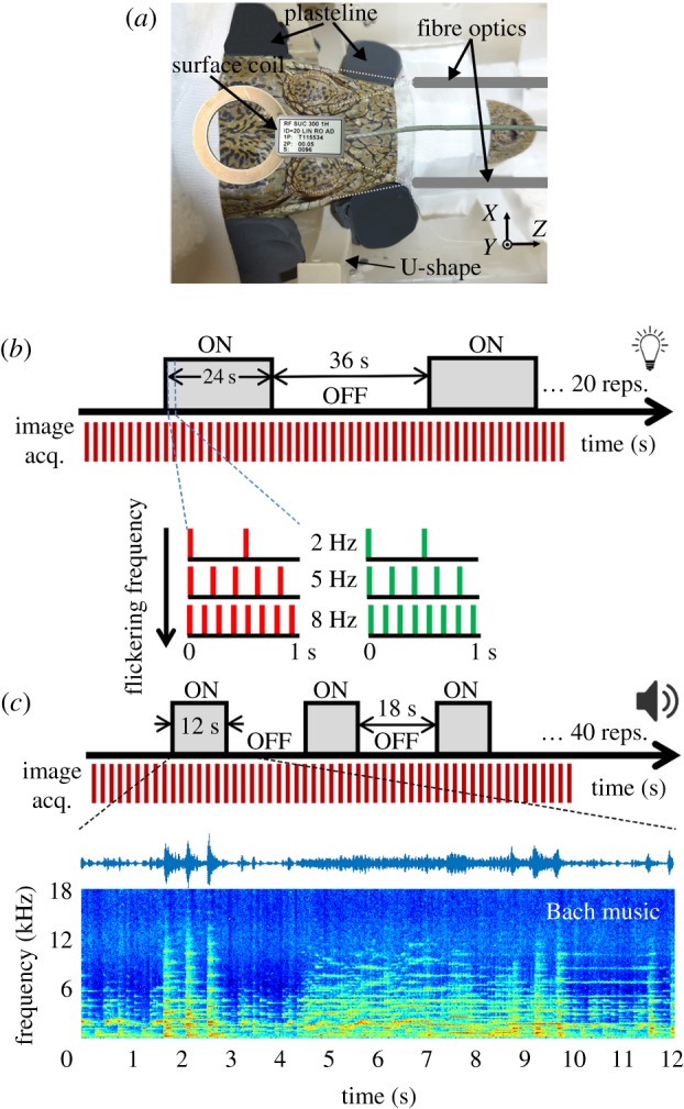
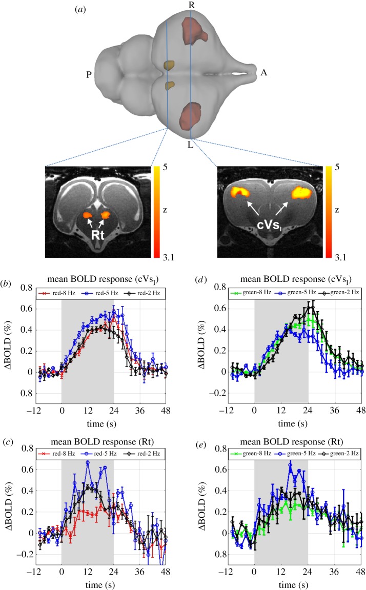
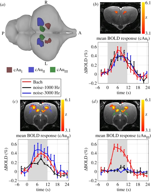
Crocodilians are important for understanding the evolutionary history of amniote neural systems as they are the nearest extant relatives of modern birds and share a stem amniote ancestor with mammals. Although the crocodilian brain has been investigated anatomically, functional studies are rare. Here, we employed functional magnetic resonance imaging (fMRI), never tested in poikilotherms, to investigate crocodilian telencephalic sensory processing. Juvenile Crocodylus niloticus were placed in a 7 T MRI scanner to record blood oxygenation level-dependent (BOLD) signal changes during the presentation of visual and auditory stimuli. Visual stimulation increased BOLD signals in rostral to mid-caudal portions of the dorso-lateral anterior dorsal ventricular ridge (ADVR). Simple auditory stimuli led to signal increase in the rostromedial and caudocentral ADVR. These activation patterns are in line with previously described projection fields of diencephalic sensory fibres. Furthermore, complex auditory stimuli activated additional regions of the caudomedial ADVR. The recruitment of these additional, presumably higher-order, sensory areas reflects observations made in birds and mammals. Our results indicate that structural and functional aspects of sensory processing have been likely conserved during the evolution of sauropsids. In addition, our study shows that fMRI can be used to investigate neural processing in poikilotherms, providing a new avenue for neurobiological research in these critical species.
Keywords: audio-visual stimulation; dorsal ventricular ridge; hierarchical processing; poikilotherms; reptile.
© 2018 The Author(s).
Conflict of interest statement
We declare we have no competing interests.
Figures
References
-
- Güntürkün O, Stacho M, Ströckens F. 2017. The brains of reptiles and birds. In Evolution of nervous systems (ed. Kaas J.), pp. 171–221. Amsterdam, Netherlands: Elsevier.
-
- Janke A, Arnason U. 1997. The complete mitochondrial genome of Alligator mississippiensis and the separation between recent archosauria (birds and crocodiles). Mol. Biol. Evol. 14, 1266–1272. (doi:10.1093/oxfordjournals.molbev.a025736) - DOI - PubMed
-
- Bird CM, Berens SC, Horner AJ, Franklin A. 2014. Categorical encoding of color in the brain. Proc. Natl Acad. Sci. USA 111, 4590–4595. (doi:10.1073/pnas.1315275111) - DOI - PMC - PubMed
-
- Bosman CA, Aboitiz F. 2015. Functional constraints in the evolution of brain circuits. Front. Neurosci. 9, 303 (doi:10.3389/fnins.2015.00303) - DOI - PMC - PubMed
-
- Jarvis ED. 2009. Evolution of the pallium in birds and reptiles. In Encyclopedia of neuroscience (eds Binder MD, Hirokawa N, Windhorst U), pp. 1390–1400. Berlin, Heidelberg, Germany: Springer Berlin Heidelberg.
Publication types
MeSH terms
Associated data
LinkOut - more resources
Full Text Sources
Other Literature Sources
Medical