. 2018 Apr 25;10(438):eaal1803.
doi: 10.1126/scitranslmed.aal1803.

Targeting protein biotinylation enhances tuberculosis chemotherapy

Affiliations

Targeting protein biotinylation enhances tuberculosis chemotherapy

Divya Tiwari et al. Sci Transl Med. .

Abstract

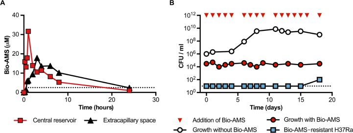
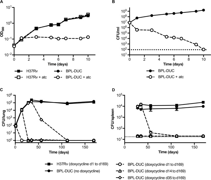
Successful drug treatment for tuberculosis (TB) depends on the unique contributions of its component drugs. Drug resistance poses a threat to the efficacy of individual drugs and the regimens to which they contribute. Biologically and chemically validated targets capable of replacing individual components of current TB chemotherapy are a major unmet need in TB drug development. We demonstrate that chemical inhibition of the bacterial biotin protein ligase (BPL) with the inhibitor Bio-AMS (5'-[N-(d-biotinoyl)sulfamoyl]amino-5'-deoxyadenosine) killed Mycobacterium tuberculosis (Mtb), the bacterial pathogen causing TB. We also show that genetic silencing of BPL eliminated the pathogen efficiently from mice during acute and chronic infection with Mtb Partial chemical inactivation of BPL increased the potency of two first-line drugs, rifampicin and ethambutol, and genetic interference with protein biotinylation accelerated clearance of Mtb from mouse lungs and spleens by rifampicin. These studies validate BPL as a potential drug target that could serve as an alternate frontline target in the development of new drugs against Mtb.


Figures

Fig. 3
Fig. 3
Activity of Bio-AMS in a hollow fiber culture system. (A) PK profile of Bio-AMS in a hollow fiber culture system. Bio-AMS concentrations were measured in samples taken from the central reservoir and the extracapillary space of the device. The dotted line represents the MIC90 of Bio-AMS for the Mtb H37Ra strain. (B) Impact of Bio-AMS on viability of the Mtb H37Ra strain in a hollow fiber culture system. The dotted line indicates the lower limit of detection.
Fig. 1
Fig. 1
Chemical inactivation of BPL kills Mtb and prevents growth in mouse macrophages. (A) Impact of Bio-AMS on viability of Mtb in standard liquid culture. The dashed line indicates the limit of detection. The frontline anti-TB drug isoniazid was used as a control. (B) Impact of Bio-AMS on growth of Mtb cultured on different carbon sources. Relative growth was calculated by dividing OD580 (optical density at 580 nm) of the culture treated with Bio-AMS by the OD580 of the culture in the absence of Bio-AMS. (C) Impact of Bio-AMS on intracellular Mtb in mouse macrophages. Mouse bone marrow–derived macrophages were infected with Mtb in vitro, and the culture was treated with Bio-AMS or dimethyl sulfoxide (DMSO) vehicle 24 hours after infection. Data are representative of two independent experiments each with three replicates and are means ± SEM (**P < 0.01, Student’s t test).
Fig. 2
Fig. 2
Emergence of Bio-AMS–resistant Mtb strains. (A) Frequency of the emergence of spontaneous resistance in Mtb cultured on standard solid medium in the presence of Bio-AMS at concentrations of 10×, 25×, and 50× the MIC. Isoniazid was used as a control. (B) Activity of Bio-AMS against Mtb strain H37Rv transformed with a multicopy plasmid expressing the Mtb protein Rv3406. The Mtb H37Rv strain contained either the vector control (pTE-mcs) or the Rv3406 expression plasmid pGMEHPtb38- rv3406. One spontaneously resistant isolate (Bio-AMS-5R1) was included as a control. (C) Saturation curve used to determine the kinetic parameters for Bio-AMS oxidation by the Mtb protein Rv3406. Left: Data of initial velocity (v0) versus concentration of Bio-AMS were fitted by nonlinear regression to the Michaelis-Menten equation. Right: Time course in milliabsorbance units (mAU) showing Rv3406-catalyzed formation of UV active metabolite 3 at 254 nm, as monitored by high-performance liquid chromatography.
Fig. 4
Fig. 4
Effect of depleting BPL on Mtb in vitro and in vivo in mice. (A and B) Impact of BPL depletion on growth (A) and survival (B) of the Mtb BPL-DUC strain in standard liquid culture medium. Growth and survival were monitored using measurements of OD and counting of CFU, respectively. (C and D) CFU isolated from mouse lungs (C) and mouse spleens (D) after infection of mice with the Mtb H37Rv strain or the Mtb mutant BPL-DUC strain. Mice infected with the Mtb H37Rv strain received doxycycline. Data are representative of three (A and B) or two (C and D) independent experiments each with three (A and B) or four (C and D) samples per time point. Data are means ± SEM.
Fig. 5
Fig. 5
Bio-AMS enhances the activity of rifampicin and ethambutol in vitro. (A to E) Impact of Bio-AMS on the activities of the TB drugs rifampicin (A and D), ethambutol (B and E), and isoniazid (C). The Mtb H37Rv strain was grown in standard liquid culture with Bio-AMS (1 µM in DMSO) or DMSO alone for 3 days, after which the bacteria were exposed to the other anti-TB drugs. Data are representative of two independent experiments with three samples per time point. Data are means ± SEM.
Fig. 6
Fig. 6
Partial inhibition of biotin synthesis increases susceptibility of Mtb to rifampicin in mice. (A) Susceptibilities of the Mtb H37Rv strain and the Mtb mutant bioA TetON-1 strain to rifampicin in biotin-free medium with and without atc. (B) Number of CFU isolated from the lungs of mice infected with the bioA TetON-1 Mtb strain. Mice were fed either doxycycline-containing (blue and purple) or doxycycline-free (gray and red) chow throughout the experiment. Rifampicin was administered 3 weeks after initial infection to one group of mice that received doxycycline (purple) and one group that had not received doxycycline (red). All samples were collected at 4 or 8 weeks after treatment with rifampicin was initiated. (C) Number of CFU isolated from the spleens of mice described in (B). Data in (B) and (C) are for n = 8 mice for each time point. Data are means ± SEM (*P < 0.05, Mann-Whitney test).

References

    1. W. H. Organization , Antimicrobial Resistance: Global Report on Surveillance (World Health Organization, 2014).
    1. Moliva J. I., Turner J., Torrelles J. B., Prospects in Mycobacterium bovis Bacille Calmette et Guérin (BCG) vaccine diversity and delivery: Why does BCG fail to protect against tuberculosis? Vaccine 33, 5035–5041 (2015). - PMC - PubMed
    1. Draper P., The outer parts of the mycobacterial envelope as permeability barriers. Front. Biosci.3: D1253–D1261 (1998). - PubMed
    1. Morris R. P., Nguyen L., Gatfield J., Visconti K., Nguyen K., Schnappinger D., Ehrt S., Liu Y., Heifets L., Pieters J., Schoolnik G., Thompson C. J., Ancestral antibiotic resistance in Mycobacterium tuberculosis. Proc. Natl. Acad. Sci. U.S.A. 102, 12200–12205 (2005). - PMC - PubMed
    1. Nguyen L., Pieters J., Mycobacterial subversion of chemotherapeutic reagents and host defense tactics: Challenges in tuberculosis drug development. Annu. Rev. Pharmacol. Toxicol. 49, 427–453 (2009). - PubMed

Publication types

LinkOut - more resources