. 2018 Jun 8;293(23):9090-9100.
doi: 10.1074/jbc.RA117.000953. Epub 2018 Apr 25.

Insights into the binding behavior of native and non-native cytochromes to photosystem I from Thermosynechococcus elongatus

Affiliations

Insights into the binding behavior of native and non-native cytochromes to photosystem I from Thermosynechococcus elongatus

Adrian Kölsch et al. J Biol Chem. .

Abstract

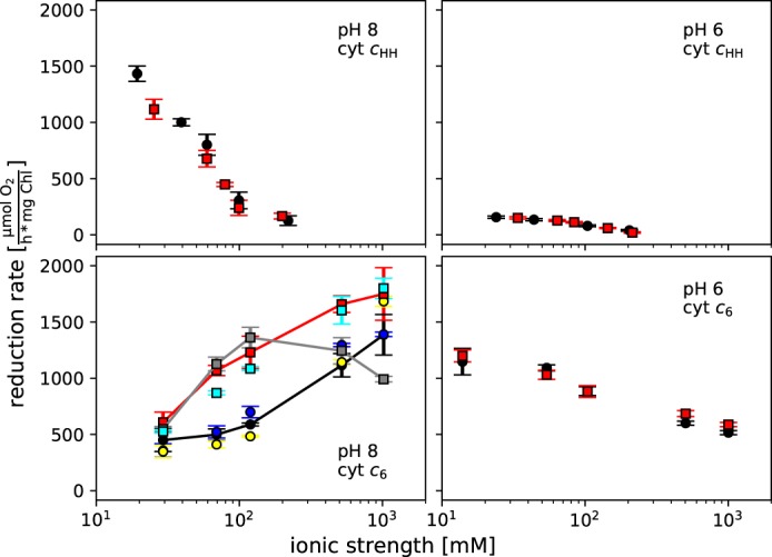
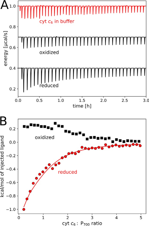
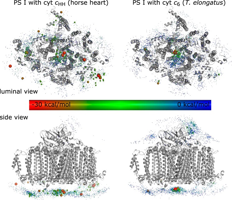
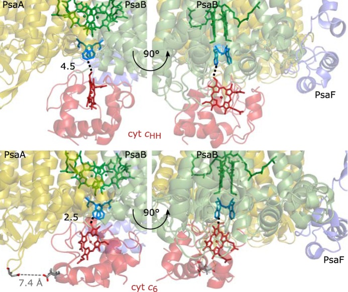
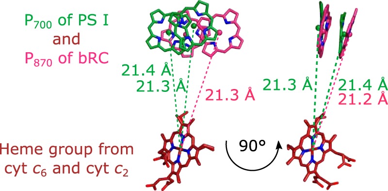
The binding of photosystem I (PS I) from Thermosynechococcus elongatus to the native cytochrome (cyt) c6 and cyt c from horse heart (cyt cHH) was analyzed by oxygen consumption measurements, isothermal titration calorimetry (ITC), and rigid body docking combined with electrostatic computations of binding energies. Although PS I has a higher affinity for cyt cHH than for cyt c6, the influence of ionic strength and pH on binding is different in the two cases. ITC and theoretical computations revealed the existence of unspecific binding sites for cyt cHH besides one specific binding site close to P700 Binding to PS I was found to be the same for reduced and oxidized cyt cHH Based on this information, suitable conditions for cocrystallization of cyt cHH with PS I were found, resulting in crystals with a PS I:cyt cHH ratio of 1:1. A crystal structure at 3.4-Å resolution was obtained, but cyt cHH cannot be identified in the electron density map because of unspecific binding sites and/or high flexibility at the specific binding site. Modeling the binding of cyt c6 to PS I revealed a specific binding site where the distance and orientation of cyt c6 relative to P700 are comparable with cyt c2 from purple bacteria relative to P870 This work provides new insights into the binding modes of different cytochromes to PS I, thus facilitating steps toward solving the PS I-cyt c costructure and a more detailed understanding of natural electron transport processes.

Keywords: complex; crystallography; cytochrome c; docking; photosynthesis; photosystem I.


Conflict of interest statement

The authors declare that they have no conflicts of interest with the contents of this article

Figures

Figure 1.
Figure 1.
Oxygen reduction rates of PS I with 16 μm cyt cHH (top) and cyt c6 (bottom) at pH 8 (left) and pH 6 (right) as a function of ionic strength. Monovalent (NaCl; black) and divalent (MgCl2; red) cations are depicted as circles and squares, respectively. For cyt c6, pH 8 (bottom, left) differences between the applied salts become prominent. Therefore, a further differentiation of the salts is shown: NaCl (black), Na2SO4 (yellow), NH4Cl (blue), MgCl2 (red), CaCl2 (gray), and MgSO4 (cyan). NaCl, MgCl2, and CaCl2 are connected by a line in their corresponding color. All measurements were performed in either 25 mm Tricine-NaOH, pH 8, or 5 mm MES-NaOH, pH 6, with 2 mm ascorbic acid and 300 μm methyl viologen at 20 °C. The concentration of buffer ions and counterions, which contribute to the ionic strength, was calculated using the Henderson–Hasselbalch equation with a pKa of 8.2 and 6.2 for Tricine and MES buffers, respectively. Standard deviations (error bars) were determined from three to nine independent measurements.
Figure 2.
Figure 2.
Isothermal titration calorimetry of PS I with cyt cHH. A, thermogram for exemplary background measurements (top) and oxidized (middle) and reduced (bottom) proteins. B, integrated heats of titrations after background subtraction in the presence (red) or absence (black) of 5 mm ascorbate. High cyt cHH:P700 ratios are omitted for a better overview. Fits (top) and residuals (bottom) are shown for one set of binding sites with n = 1.0 (dashed line) and for one set of binding sites with n = 1.5 (solid line). Parameters obtained from the models are shown in Table 2. Measurements were performed at 20 °C in 25 mm Tricine buffer, pH 8.0, with 25 mm NaCl and 0.02% DDM. Each titration step consisted of a 5-μl injected volume from 1 mm cyt cHH.
Figure 3.
Figure 3.
Isothermal titration calorimetry of PS I with cyt c6. A, thermogram for exemplary background measurements (top, red) and oxidized (middle) and reduced (bottom) proteins. B, integrated heats of titrations after background subtraction in the presence (red; reduced) or absence (black; oxidized) of 5 mm ascorbate. The fit of the data for reduced cyt c6 is shown for one set of binding sites with n = 1.0. Parameters obtained from the model are shown in Table 2. After substraction of the heat of dilution, the data for oxidized cyt c6 converge to negative values at high cyt c6:PS I ratio (−0.1 kcal/mol; not shown) and are thus not analyzed by a model. Measurements were performed at 20 °C in 25 mm Tricine-NaOH, pH 8.0, with 25 mm NaCl and 0.02% DDM. Each titration step consisted of a 5-μl injected volume from 1 mm cyt c6.
Figure 4.
Figure 4.
Molecular docking simulation of monomeric PS I with cyt cHH (left) and cyt c6 (right). Each sphere represents the position of a docked cyt c. The binding energy, calculated by pyDock, is highlighted by a color code. Docking states with less than −20 kcal/mol are highlighted by an increased sphere size.
Figure 5.
Figure 5.
Potential cyt cHH (top)– and cyt c6 (bottom)–binding site of PS I. Shown are the docking sites that most likely resemble the specific cyt c–binding site of PS I. The heme group (red) of cyt cHH and cyt c6 points toward the luminal tryptophan residues Trp-A655 and Trp-B631 (blue) and P700 (green) of PS I. The distances between the heme groups and the closest tryptophan are highlighted by a black dotted line. Cyt c6 does not interact with PsaF (purple) but is close to the luminal loop of PsaA (yellow). The carboxyl group of Glu-34 from cyt c6 is at a distance of 7.4 Å from the carboxyl group of Asp-628 from PsaA (gray dotted line).
Figure 6.
Figure 6.
Superposition of the potential cyt c6–binding site to the known cyt c2–binding site of the bRC from R. sphaeroides (Protein Data Bank code 1l9b (34)). The superposition was achieved by aligning the heme groups. The right view is rotated by 90° with respect to the left view. The distances of the heme iron from cyt c6 to the Mg2+ ions of P700 are 21.4 and 21.3 Å, respectively. These distances are identical to the distances between the heme iron of cyt c2 and the Mg2+ ions of P870 (pink) from bRC (21.3 and 21.2 Å, respectively).

References

    1. Stieger K. R., Feifel S. C., Lokstein H., Hejazi M., Zouni A., and Lisdat F. (2016) Biohybrid architectures for efficient light-to-current conversion based on photosystem I within scalable 3D mesoporous electrodes. J. Mater. Chem. A 4, 17009–17017 10.1039/C6TA07141D - DOI
    1. Stieger K. R., Ciornii D., Kölsch A., Hejazi M., Lokstein H., Feifel S. C., Zouni A., and Lisdat F. (2016) Engineering of supramolecular photoactive protein architectures: the defined co-assembly of photosystem I and cytochrome c using a nanoscaled DNA-matrix. Nanoscale 8, 10695–10705 10.1039/C6NR00097E - DOI - PubMed
    1. Lubner C. E., Applegate A. M., Knörzer P., Ganago A., Bryant D. A., Happe T., and Golbeck J. H. (2011) Solar hydrogen-producing bionanodevice outperforms natural photosynthesis. Proc. Natl. Acad. Sci. 108, 20988–20991 10.1073/pnas.1114660108 - DOI - PMC - PubMed
    1. Nowaczyk M. M., and Plumeré N. (2016) Short circuit at the chlorophyll. Nat. Chem. Biol. 12, 990–991 10.1038/nchembio.2240 - DOI - PubMed
    1. Jordan P., Fromme P., Witt H. T., Klukas O., Saenger W., and Krauss N. (2001) Three-dimensional structure of cyanobacterial photosystem I at 2.5 Å resolution. Nature 411, 909–917 10.1038/35082000 - DOI - PubMed

Publication types