

Surgical Management of a Giant Pheochromocytoma
- PMID: 29695582
- PMCID: PMC6000789
- DOI: 10.21873/invivo.11297
Surgical Management of a Giant Pheochromocytoma
Abstract
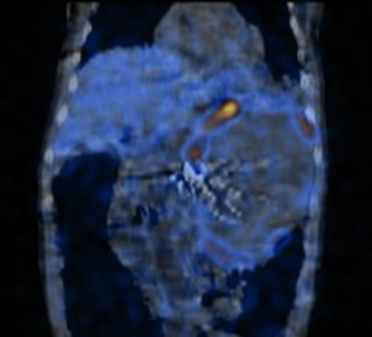
Pheochromocytomas are rare tumors arising from chromaffin cells of the adrenal medulla and extramedullary sympathetic ganglia. The incidence of asymptomatic disease is rising due to increased detection rates from widespread use of computed tomography. We describe a case of one of the largest documented pheochromocytomas resected in the United States, an 18-cm tumor in a patient who presented with exertional dyspnea, abdominal pain, constipation, weight loss, and intermittent hypertension. After biochemical and appropriate imaging workup, the patient underwent an open resection of the mass.
Keywords: Pheochromocytoma; adrenal; endocrine; kidney; paraganglionoma.
Copyright© 2018, International Institute of Anticancer Research (Dr. George J. Delinasios), All rights reserved.
Figures


References
-
- Stein PP, Black HR. A simplified diagnostic approach to pheochromocytoma. A review of the literature and report of one institution’s experience. Medicine. 1991;70(1):46–66. - PubMed
-
- Neumann HP, Berger DP, Sigmund G, Blum U, Schmidt D, Parmer RJ, Volk B, Kirste G. Pheochromocytomas, multiple endocrine neoplasia type 2, and von Hippel-Lindau disease. N Engl J Med. 1993;329(21):1531–1538. - PubMed
-
- Walther MM, Herring J, Enquist E, Keiser HR, Linehan WM. von Recklinghausen’s disease and pheochromocytomas. J Urol. 1999;162(5):1582–1586. - PubMed
-
- Quan-Yang D, Yeh MW. The adrenal glands. Chapter 39: In: Textbook of Surgery: The Biological Basis of Modern Surgical Practice. Philadelphia: W.B: Saunders. 1997:pp. 1016–017.
Publication types
MeSH terms
LinkOut - more resources
Full Text Sources
Other Literature Sources
Medical