. 2018 May 15;115(20):5235-5240.
doi: 10.1073/pnas.1722151115. Epub 2018 Apr 25.

CRISPR/Cas9-mediated genome editing in a reef-building coral

Affiliations

CRISPR/Cas9-mediated genome editing in a reef-building coral

Phillip A Cleves et al. Proc Natl Acad Sci U S A. .

Abstract

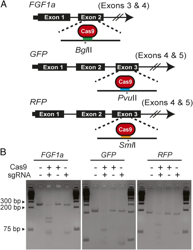
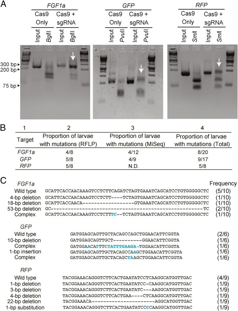
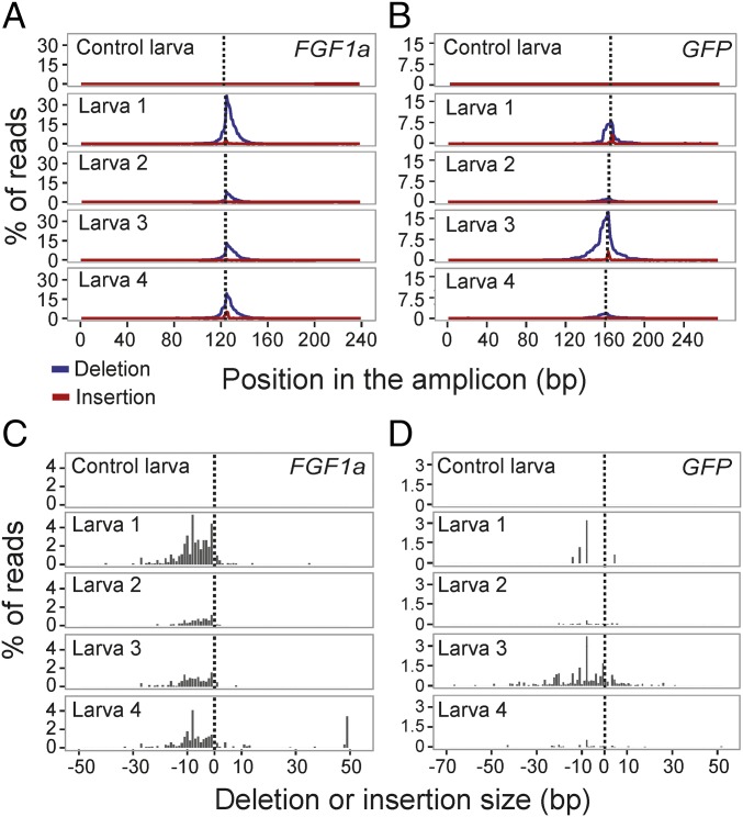
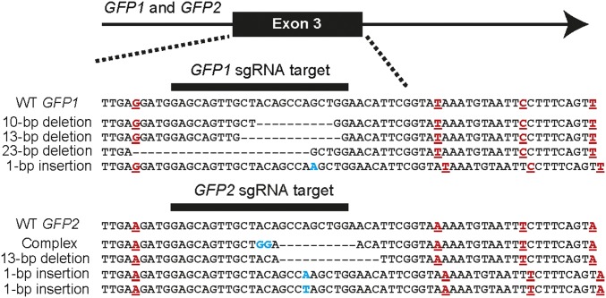
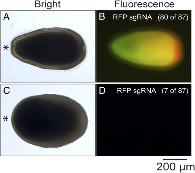
Reef-building corals are critically important species that are threatened by anthropogenic stresses including climate change. In attempts to understand corals' responses to stress and other aspects of their biology, numerous genomic and transcriptomic studies have been performed, generating a variety of hypotheses about the roles of particular genes and molecular pathways. However, it has not generally been possible to test these hypotheses rigorously because of the lack of genetic tools for corals. Here, we demonstrate efficient genome editing using the CRISPR/Cas9 system in the coral Acropora millepora We targeted the genes encoding fibroblast growth factor 1a (FGF1a), green fluorescent protein (GFP), and red fluorescent protein (RFP). After microinjecting CRISPR/Cas9 ribonucleoprotein complexes into fertilized eggs, we detected induced mutations in the targeted genes using changes in restriction-fragment length, Sanger sequencing, and high-throughput Illumina sequencing. We observed mutations in ∼50% of individuals screened, and the proportions of wild-type and various mutant gene copies in these individuals indicated that mutation induction continued for at least several cell cycles after injection. Although multiple paralogous genes encoding green fluorescent proteins are present in A. millepora, appropriate design of the guide RNA allowed us to induce mutations simultaneously in more than one paralog. Because A. millepora larvae can be induced to settle and begin colony formation in the laboratory, CRISPR/Cas9-based gene editing should allow rigorous tests of gene function in both larval and adult corals.

Keywords: Acropora millepora; CRISPR/Cas9; coral; genome editing.


Conflict of interest statement

The authors declare no conflict of interest.

Figures

Fig. 1.
Fig. 1.
Design and activity in vitro of sgRNAs targeting A. millepora genes. (A) sgRNAs targeting exon 2 (of at least four) of FGF1a, exon 3 (of five) of GFP genes, and exon 3 (of five) of RFP genes were designed to induce double-strand breaks near endogenous restriction-enzyme sites that could be used to detect induced mutations. Colored bars, approximate locations of the sgRNA-binding sites; asterisks, predicted Cas9 cleavage sites and the nearby restriction sites. (B) Digestion in vitro of FGF1a, GFP, or RFP target DNA (SI Appendix, SI Materials and Methods) incubated with Cas9 protein, the appropriate sgRNA (as transcribed in vitro from the pDR274-based construct), or both. Fragments were analyzed by gel electrophoresis; outside lanes of each gel show molecular-size markers.
Fig. 2.
Fig. 2.
Efficient CRISPR/Cas9-mediated genome editing in injected A. millepora embryos. (A) Genomic DNA from individual larvae that had been injected with Cas9 protein alone or with the sgRNA/Cas9 protein complexes for one of the target genes was amplified by PCR, digested with the appropriate restriction enzyme, and analyzed by gel electrophoresis. Each pair of interior lanes represents one larva; the outside lanes show molecular-size markers. Arrows indicate the incompletely digested DNA. (B) Proportions of injected larvae with mutations as determined by restriction digestion (RFLP, as in A) or MiSeq amplicon sequencing (SI Appendix, SI Materials and Methods). The total numbers of larvae tested by each method and the numbers found to be carrying mutations are indicated. N.D., not determined. Columns are numbered for ease of reference in the text. (C) Varieties of specific mutations in individual larvae. For each gene, genomic DNA was PCR-amplified and cloned from one injected larva that appeared from RFLP analysis (A and B) to harbor mutations, the indicated numbers of clones were Sanger-sequenced, and the sequences were aligned. Base-pair changes are shown in blue and deletions by dashes; the numbers of wild-type and variant sequences observed are indicated. Note that for GFP and RFP, the sequences shown could be from more than one paralog in each case (see text).
Fig. 3.
Fig. 3.
Mutational spectra in CRISPR/Cas9-injected animals as determined by MiSeq amplicon sequencing. For each gene, one control larva (only Cas9 protein injected) and the four sgRNA/Cas9-injected larvae determined by MiSeq sequencing to contain mutations (Fig. 2B, column 3) were analyzed further. (A and B) The percentages of reads with a deleted (blue lines) or inserted (red lines) base at each nucleotide position. Dotted lines, expected Cas9 cut sites. Note that for GFP, the method of analysis used here does not resolve the paralogous copies of the gene (SI Appendix, SI Materials and Methods). (C and D) The percentages of reads with deletions (Left of dotted line) or insertions (Right of dotted line) of various sizes in the same larvae as shown in A and B.
Fig. 4.
Fig. 4.
Editing of two GFP paralogs in a single larva with a single sgRNA. The sequences from larva 4 (Fig. 3B) were clustered into eight groups using the single-nucleotide differences within an 83-bp intronic region (SI Appendix, Fig. S1). Shown are representative mutant alleles that fell into groups 1 and 6 (SI Appendix, Fig. S1), which can be assigned with confidence to GFP1 and GFP2, respectively (SI Appendix, SI Materials and Methods). Dashes, deleted nucleotides; blue lettering, inserted or altered nucleotides; red lettering, positions at which the GFP1 and GFP2 alleles in this larva differed within this 64-bp segment. (Note that only two of these positions are among those diagnostic for GFP1 vs. GFP2: cf. SI Appendix, Fig. S1.)
Fig. 5.
Fig. 5.
GFP (green) and RFP (red) fluorescence in larvae that had been injected with RFP sgRNA/Cas9 complexes. Larvae were observed at 5 d postfertilization; bright-field and merged GFP- and RFP-fluorescence images are shown for each larva. (A and B) Most injected larvae showed fluorescence patterns indistinguishable from those of wild type. (C and D) Some injected larvae showed neither GFP nor RFP fluorescence. Asterisk (*), oral end of larva. (Scale bar, 200 µm.)

References

    1. Moberg F, Folke C. Ecological goods and services of coral reef ecosystems. Ecol Econ. 1999;29:215–233.
    1. Hughes TP, et al. Coral reefs in the Anthropocene. Nature. 2017;546:82–90. - PubMed
    1. Muscatine L, Cernichiari E. Assimilation of photosynthetic products of zooxanthellae by a reef coral. Biol Bull. 1969;137:506–523. - PubMed
    1. Muscatine L, Falkowski PG, Porter JW, Dubinsky Z. Fate of photosynthetic fixed carbon in light- and shade-adapted colonies of the symbiotic coral Stylophora pistillata. Proc R Soc B. 1984;222:181–202.
    1. Davy SK, Allemand D, Weis VM. Cell biology of cnidarian-dinoflagellate symbiosis. Microbiol Mol Biol Rev. 2012;76:229–261. - PMC - PubMed

Publication types

Substances

LinkOut - more resources