Comparative Study
. 2018 May;20(5):553-564.
doi: 10.1038/s41556-018-0089-0. Epub 2018 Apr 25.

TFAP2C regulates transcription in human naive pluripotency by opening enhancers

Affiliations
Comparative Study

TFAP2C regulates transcription in human naive pluripotency by opening enhancers

William A Pastor et al. Nat Cell Biol. 2018 May.

Abstract

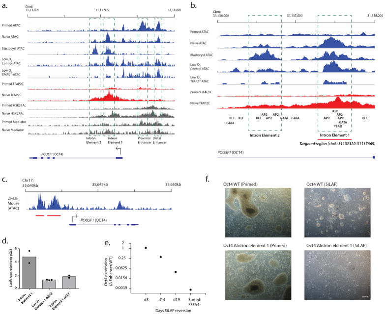
Naive and primed pluripotent human embryonic stem cells bear transcriptional similarity to pre- and post-implantation epiblast and thus constitute a developmental model for understanding the pluripotent stages in human embryo development. To identify new transcription factors that differentially regulate the unique pluripotent stages, we mapped open chromatin using ATAC-seq and found enrichment of the activator protein-2 (AP2) transcription factor binding motif at naive-specific open chromatin. We determined that the AP2 family member TFAP2C is upregulated during primed to naive reversion and becomes widespread at naive-specific enhancers. TFAP2C functions to maintain pluripotency and repress neuroectodermal differentiation during the transition from primed to naive by facilitating the opening of enhancers proximal to pluripotency factors. Additionally, we identify a previously undiscovered naive-specific POU5F1 (OCT4) enhancer enriched for TFAP2C binding. Taken together, TFAP2C establishes and maintains naive human pluripotency and regulates OCT4 expression by mechanisms that are distinct from mouse.


Figures

Figure 1
Figure 1. Determination of regulatory elements specific to the naïve and primed states in humans
a,b Percentage of the time a gene whose transcriptional start site is a given distance from a naïve-specific ATAC-peak (a) or primed-specific ATAC-peak (b) is upregulated or downregulated in the naïve state. c,d Many naïve-specific ATAC-seq peaks appear proximal to KLF5 (c), a gene highly upregulated in the naïve state, while primed-specific ATAC-peaks are present near the primed-specific ZIC genes. Note the strong enrichment of H3K27Ac near ATAC-seq peaks. H3K4me3 and H3K27Ac ChIP-seq data come from published sources,. e, Metaplot of H3K27Ac and Mediator over naïve-specific (left) and primed specific (right) ATAC-peaks. f,g Most statistically significant transcription factor binding motifs enriched in naïve-specific (f) or primed-specific (g) ATAC peaks were calculated using a cumulative binomial distribution. Pooled data from 4 naïve and 4 primed biological replicates were used.
Figure 2
Figure 2. Most naïve-specific ATAC-peaks are present in other naïve human cells and the human embryo
a, Normalized ATAC-seq reads from the human blastocyst plotted relative to Naïve-specific and primed-specific peaks. Note far greater enrichment over naïve-specific peaks. b, Blastocyst ATAC-seq plotted relative to all Naïve-specific ATAC peaks. Note enrichment over almost all naïve-specific peaks, indicating that they are open in the blastocyst. c, Most Naïve-specific ATAC-seq peaks overlap with a blastocyst-ATAC peak, but most primed-specific peaks do not. d,e ATAC-seq signal for primed hESCs, naïve hESCs and blastocyst in the viscinity of NANOG (d) and GATA3 (e). Peak height is normalized to total number of reads in each sample. f,g Metaplot of ATAC-seq read density over the gene bodies of 100 genes most highly specific to trophoblast or epiblast, as defined from single-cell RNA-seq data in human, as well as all genes. h, Venn diagram showing overlap of all ATAC-seq peaks in blastocyst, naïve hESCs and primed hESCs. i, Enrichment of GATA, AP2, KLF and OCT-SOX motifs in each set identified in part e. Note enrichment of AP2 and KLF motifs in both blastocyst and naïve hESCs, stronger enrichment of GATA in blastocyst and stronger enrichment of OCT-SOX in ESCs.
Figure 3
Figure 3. TFAP2C is highly enriched over naïve-specific open chromatin in humans
a, TFAP2C is highly expressed in naïve cells, both relative to other AP2 transcription factors and relative to primed cells. Mean and standard deviation are shown, with dots representing each replicate (n=4 independent experiments). b, TFAP2C protein is highly upregulated in the naïve-pluripotent state. Data represent 1 out of 5 independent experiments with similar results. c, Strong co-enrichment of TFAP2C with naïve-specific ATAC peaks at the CBFA2T2 locus. d, Global enrichment of TFAP2C relative to the summits of different categories of naïve-specific ATAC peaks. TFAP2C is enriched over naïve-specific ATAC peaks, especially those with AP2 motifs. e,f TFAP2C is strongly enriched over Naïve-specific ATAC peak summits compared with enrichment over regions that show ATAC enrichment in both naïve and primed cells (Naïve-primed Intersect) (e), even though both peak sets show similar ATAC enrichment (f). Uncropped Western blot images are available in Supplementary Figure 9. Source data for a is available in Supplementary Table 8.
Figure 4
Figure 4. TFAP2C−/− cells differentiate in naïve media
a, TFAP2C−/− hESCs, self-renew in primed conditions but differentiate and fail to self-renew upon treatment in naïve (5iLAF) media. Scale bars indicate 100 μm. Data represent 1 out of 4 independent experiments with similar results. b, Western blot of TFAP2C upon culture in primed or 5iLAF conditions. TFAP2C is strongly induced within three days of treatment with 5iLAF. Data represent 1 out of 2 independent experiments with similar results. c, Western blot for TFAP2C after five days of 5iLAF culture. TFAP2C is absent from TFAP2C−/− deficient lines. d, ATAC-seq openness of Naïve, d5 5iLAF WT and TFAP2C−/−, and primed cells over naïve-specific ATAC peaks. Note that after five days of reversion, substantial opening of the naïve-specific ATAC peaks has already occurred, but not in the TFAP2C−/−cells. e, TFAP2C ChIP enrichment shown over naïve and d5 5iLAF samples. ChIP input for each set is shown as a dashed line. f, Western blot for OCT4 and NANOG in control and TFAP2C−/− cells after five days of culture in 5iLAF. Quantitation normalized to histone below. g, Western Blot for SOX1 and PAX6 in control and TFAP2C−/− cells. h, Relative RPKM of pluripotency and neural markers in RNA-seq. Data is from n=3 WT and n=4 TFAP2C−/− independent biological replicates (mean +/− s.e). i,j, Immunofluorescent staining for TFAP2C, OCT4 (i) and PAX6 (j) in control and TFAP2C−/− cells. Scale bar indicates 20 μm. k,l Fold enrichment for AP2 motifs in the specified peak sets in humans (k) and mouse (l). Asterix indicates no enrichment. Although AP2 motifs are enriched in naïve-specific peaks in both species, the enrichment is much stronger in the human naïve-specific set. m, Expression of key pluripotency markers in WT, Tfap2c−/−, and Tfap2a−/− Tfap2c−/− cells in 2i+LIF conditions. n=4 biological replicates in for Tfap2c−/− and controls and n=6 biological replicates for Tfap2a−/− Tfap2c−/− and controls (mean +/− s.e.) n, ATAC-seq peaks specific to WT and Tfap2a−/− Tfap2c−/− were calculated and enrichment for AP2 motifs determined. Asterisk indicates no enrichment. Uncropped Western blots in Supplementary Figure 9. Source data for h and m in Supplementary Table 8.
Figure 5
Figure 5. Ectopic expression of TFAP2C partially rescues TFAP2C−/− phenotype
a, Quantitative western blot showing tunable TFAP2C induction in TFAP2C−/− background in primed conditions. b,c Western blots show rescue of OCT4 expression and SOX1 repression upon doxycycline inducible TFAP2C expression. Lysates were collected after five days of treatment with 5iLAF and the indicated concentration of doxycycline. d, Appearance of round naïve-like colonies in lines with ectopic TFAP2C expression. Scale bar indicates 100μm. Results represent 1 out of 4 independent experiments with similar results. e, Partial rescue of upregulation of naïve pluripotency factors, downregulation of primed-factors with ectopic TFAP2C expression. 1 replicate for dox induction samples and primed control, 4 for naïve samples and primed control. f, TFAP2C was ectopically expressed for the first 15 days of reversion, then removed in some cells to induce acute loss of TFAP2C. ATAC-seq plotted from these cells is plotted over naïve-specific peaks (5032 peaks), a subset that contained an AP2 motif but no KLF motif (1054 peaks), a subset that contained a KLF motif but no AP2 motif (1551 peaks) and primed-specific peaks (2562 peaks). Reduced ATAC-seq density over naïve specific peaks and increased density over primed-specific peaks, in the sample in which doxycycline had been withdrawn. Closing of naïve specific peaks is especially pronounced over the subset of peaks that contain AP2 sites but no KLF sites (AP2+ KLF-). Peaks subsets are listed in Supplementary Table 2. Uncropped Western blot images are available in Supplementary Figure 9. Source data for e is available in Supplementary Table 8
Figure 6
Figure 6. TFAP2C−/− cells survive in 5iLAF in 5% O2 conditions but do not transition to naïve state
a, Western blots for the pluripotency marker OCT4 and the neural markers SOX1 and PAX6 in WT and TFAP2C−/− cells after 5 days in 5iLAF at 5% O2. b,c Brightfield images of control and TFAP2C−/− cells in 5iLAF culture. Initially the TFAP2C−/− cells show morphology similar to what is observed in ambient oxygen concentration conditions (compare to Figure 4A). However, some colonies are observable after passaging. These colonies show a shift toward SSEA4+ (primed) state. Scale bars indicates 100μm. c, ATAC-seq data from control and TFAP2C−/− cells in 5% O2 plotted over ATAC-seq peak sets. d, Principle component analysis comparing ATAC-seq datasets generated in this work. Blue dots: after five days in 5iLAF, WT control cells show an ATAC-seq landscape part-way between primed and naïve, whereas TFAP2C−/− cells show no change toward naïve. Green dots: although TFAP2C−/− cells survive in low oxygen conditions, they have an ATAC-seq landscape much more similar to primed than naïve cells. Red dots: ectopic doxycycline dependent-expression of TFAP2C in TFAP2C−/− partially rescues the naïve-landscape, and withdrawal of doxycycline induces a shift toward primed identity. Shown for comparison are control cells reverted at the same time. e, Genes differentially regulated in naïve vs. primed hESCs are plotted. Note that genes higher expressed in naïve cells are lower expressed in TFAP2C−/−. The RPKM values correspond to SSEA4 cells in control (average of 2 biological replicates) and SSEA4+ in TFAP2C−/− (average of 3 biological replicates). Uncropped Western blot images are available in Supplementary Figure 9.
Figure 7
Figure 7. Identifying direct regulatory targets of TFAP2C
a, Percentage of the time a gene whose transcriptional start site is a given distance from a TFAP2C ChIP-seq peak is upregulated or downregulated in naïve hESCs. Notice the much weaker correspondence compared with Figure 1A, and the lack of any effect at the promoter. b, Distance of TFAP2C ChIP-seq peak to nearest promoter. c, To identify pluripotency-state genes positively or negatively regulated by TFAP2C, we identified the subset of naïve-specific genes downregulated <4 fold in TFAP2C−/− (positively related by TFAP2C) and primed-specific genes upregulated >4 fold in TFAP2C−/− (negatively regulated by TFAP2C). Because they were the predominant pluripotent populations, we compared expression of SSEA4 control cells (n=2 biological replicates) to SSEA4+ TFAP2C−/− cells (n=3 biological replicates) d, To identify TFAP2C-dependent enhancers, we identified the overlap of the naïve-specific and TFAP2C ChIP-seq peaks, then took the subset of peaks that showed >50% density reduction in TFAP2C−/− SSEA4+ as compared with control SSEA4 cells, normalized for total read depth. These were classified as TFAP2C-dependent regulatory elements. e, ATAC-seq read density over all naïve-specific ATAC-peaks, naïve-specific ATAC peaks overlapping with TFAP2C ChIP-seq peaks, and the TFAP2C-dependent regulatory element set identified in (d). Note dramatic loss of signal in TFAP2C−/− over the TFAP2C-dependent set. f, Frequency with which a gene a given distance from a TFAP2C-dependent ATAC-seq peak is positively or negatively regulated by TFAP2C. g, Distance of TFAP2C-dependent ATAC-seq peak to nearest gene. Note that the vast majority of such elements are enhancers. h, Schematic demonstrating the typical regulatory role of TFAP2C in naïve hESCs. Where TFAP2C facilitates the opening of a new enhancer, it has a positive regulatory role. Where it hones to chromatin that is already open, it has no tangible effect on transcription. i, ATAC-seq and ChIP-seq data are shown in the vicinity of naïve-pluripotency factor TFCP2L1.
Figure 8
Figure 8. A TFAP2C+ intronic enhancer of OCT4
a, Chromatin landscape of OCT4. Two putative enhancers “intron element 1” and “intron element 2” are present in blastocyst. Intron element 1 is also strongly enriched in naïve cells and lost in TFAP2C−/ −. b, The location of consensus motifs for key pre-implantation transcription factors is shown in the vicinity of Intron Elements 1 and 2. Note the clustering of AP2 sites at each element. The control low O2 track is the SSEA4 population, the TFAP2C−/− low O2 is the SSEA4+ population. The region targeted for CRISPR deletion is shown. c, ATAC-seq reads over the murine POU5F1 locus in naïve (2i+LIF) conditions. Note the absence of either intronic enhancer. d, Luciferase activity from a pGL3 construct in which WT or mutant Intron Element 1 had been cloned, normalized to signal from a pGL3 construct with no enhancer. Results are shown from two independent experiment, except for the ΔAP2 sample, for which there are n=3 replicates from two experiments. All signals were first normalized for Renilla signal. e, OCT4 expression is lost over time upon reversion of the intron element 1-deleted mutant, indicating differentiation. Sorting for SSEA4 cells in 5iLAF culture typically produces a pure population of naïve hESCs, but this population has lost OCT4 expression in the intron element 1-deleted mutant. Mean of n=2 technical replicates is shown. f, A line in which the intron element 1 is deleted appears normal in primed conditions but fails to yield naïve colonies upon reversion. Scale bar indicates 200μm. Images are representative of 3 independent reversions. Source data for d is available in Supplementary Table 8.

References

    1. Niakan KK, Han J, Pedersen RA, Simon C, Pera RA. Human pre-implantation embryo development. Development. 2012;139:829–841. doi: 10.1242/dev.060426. - DOI - PMC - PubMed
    1. Okamoto I, et al. Eutherian mammals use diverse strategies to initiate X-chromosome inactivation during development. Nature. 2011;472:370–374. doi: 10.1038/nature09872. - DOI - PubMed
    1. Petropoulos S, et al. Single-Cell RNA-Seq Reveals Lineage and X Chromosome Dynamics in Human Preimplantation Embryos. Cell. 2016;165:1012–1026. doi: 10.1016/j.cell.2016.03.023. - DOI - PMC - PubMed
    1. Sahakyan A, et al. Human Naive Pluripotent Stem Cells Model X Chromosome Dampening and X Inactivation. Cell Stem Cell. 2017;20:87–101. doi: 10.1016/j.stem.2016.10.006. - DOI - PMC - PubMed
    1. Kunath T, et al. Developmental differences in the expression of FGF receptors between human and mouse embryos. Placenta. 2014;35:1079–1088. doi: 10.1016/j.placenta.2014.09.008. - DOI - PubMed

Publication types

MeSH terms