Review
. 2018 Apr 11:12:12.
doi: 10.3389/fnsys.2018.00012. eCollection 2018.

Interfacing Graphene-Based Materials With Neural Cells

Affiliations
Review

Interfacing Graphene-Based Materials With Neural Cells

Mattia Bramini et al. Front Syst Neurosci. .

Abstract

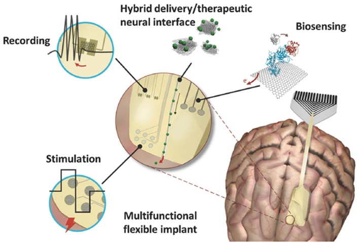
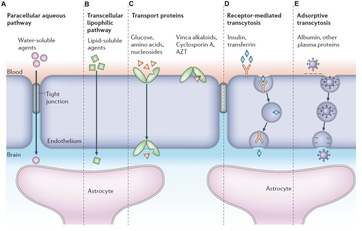
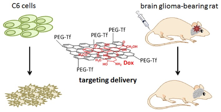
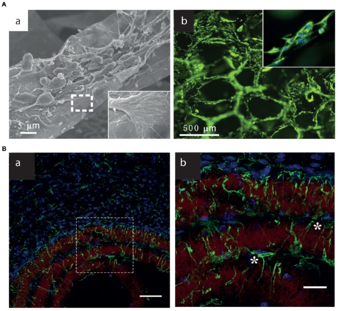
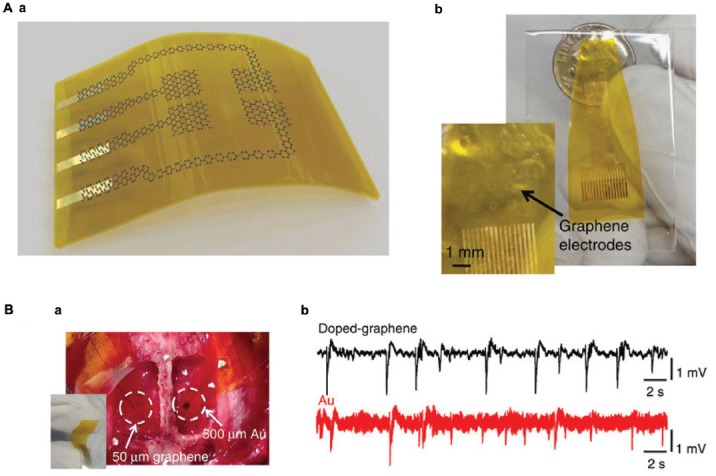
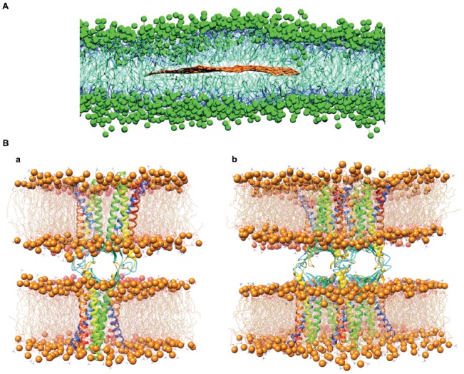
The scientific community has witnessed an exponential increase in the applications of graphene and graphene-based materials in a wide range of fields, from engineering to electronics to biotechnologies and biomedical applications. For what concerns neuroscience, the interest raised by these materials is two-fold. On one side, nanosheets made of graphene or graphene derivatives (graphene oxide, or its reduced form) can be used as carriers for drug delivery. Here, an important aspect is to evaluate their toxicity, which strongly depends on flake composition, chemical functionalization and dimensions. On the other side, graphene can be exploited as a substrate for tissue engineering. In this case, conductivity is probably the most relevant amongst the various properties of the different graphene materials, as it may allow to instruct and interrogate neural networks, as well as to drive neural growth and differentiation, which holds a great potential in regenerative medicine. In this review, we try to give a comprehensive view of the accomplishments and new challenges of the field, as well as which in our view are the most exciting directions to take in the immediate future. These include the need to engineer multifunctional nanoparticles (NPs) able to cross the blood-brain-barrier to reach neural cells, and to achieve on-demand delivery of specific drugs. We describe the state-of-the-art in the use of graphene materials to engineer three-dimensional scaffolds to drive neuronal growth and regeneration in vivo, and the possibility of using graphene as a component of hybrid composites/multi-layer organic electronics devices. Last but not least, we address the need of an accurate theoretical modeling of the interface between graphene and biological material, by modeling the interaction of graphene with proteins and cell membranes at the nanoscale, and describing the physical mechanism(s) of charge transfer by which the various graphene materials can influence the excitability and physiology of neural cells.

Keywords: blood-brain barrier; brain; computational modeling; graphene; nanomedicine; neurology; scaffolds; smart materials.


Figures

Figure 1
Figure 1
Graphene based neural interfaces for a variety of neuronal functionalities like recording, stimulation and biosensing. Modified with permission from Kostarelos et al. (2017).
Figure 2
Figure 2
Pathways across the blood-brain barrier (BBB). Modified with permission from Abbott et al. (2006).
Figure 3
Figure 3
Transferrin modified G oxide (GO) for glioma-targeted drug delivery. Modified with permission from Liu G. et al. (2013).
Figure 4
Figure 4
3D G-Scaffolds in vitro and in vivo. (A) (a) SEM images of neural stem cells (NSCs) cultured on 3D-G foams under proliferation conditions. The insets illustrate the interaction between the cell filopodia and surface. (b) Fluorescence images of NSCs cultured on 3D-G foams for 5 days. Nestin (green) is a marker for NSCs, and DAPI (blue) identifies nuclei. Modified with permission from Li N. et al. (2013). (B) (a,b) Brain astrocyte/G-scaffolds interaction and astrocyte process infiltration 3 weeks after scaffold implantation. Green: GFAP-positive astrocytes, blue: DAPI-stained nucleus, red: surface-functionalized scaffolds. (b) Detailed astrocyte morphology of the dash-box indicated area in (a). *Indicate astrocytes that bridge a gap between two scaffold layers. Scale bar, 50 (a) and 20 (b) μm. Modified with permission from Zhou et al. (2016).
Figure 5
Figure 5
Graphene electrodes for in vivo recording. (A) (a) Schematic illustration of a flexible G neural electrode array. (b) Photograph of a 16-electrode transparent array. The electrode size is 300 × 300 μm2. (B) (a) Photograph of a 50 × 50 μm2 single-G electrode placed on the cortical surface of the left hemisphere and a 500 × 500 μm2 single-Au electrode placed on the cortical surface of the right hemisphere. (b) Interictal-like spiking activity recorded by 50 × 50 μm2 doped-G and Au electrodes. Recordings with doped-G electrodes are five- to sixfold less noisy compared with the ones with same size Au electrode. Modified with permission from Kuzum et al. (2014).
Figure 6
Figure 6
Graphene interaction with biomembranes. (A) Equilibrated superstructure of a graphene sheet inside the phospholipid bilayer formed by 1-palmitoyl-2-oleoyl-sn-glycero-3-phosphocholine (POPC) lipids. Polar heads of the POPC lipids are depicted as green beads, hydrophobic hydrocarbon chains as thick blue lines; the graphene sheet is shown with brown lines (water molecules not shown; modified with permission from Titov et al., 2010). (B) The structure of single (a) and double (b) Cldn15-based paracellular pores, after the respective equilibration protocols. Protomers are shown as ribbons. Each cis dimer is embedded in a hexagonal POPC bilayer, shown as wire structures with phosphorus atoms as spheres. Modified with permission from Alberini et al. (2017).

References

    1. Abbott N. J. (2013). Blood-brain barrier structure and function and the challenges for CNS drug delivery. J. Inherit. Metab. Dis. 36, 437–449. 10.1007/s10545-013-9608-0 - DOI - PubMed
    1. Abbott N. J., Patabendige A. A., Dolman D. E., Yusof S. R., Begley D. J. (2010). Structure and function of the blood-brain barrier. Neurobiol. Dis. 37, 13–25. 10.1016/j.nbd.2009.07.030 - DOI - PubMed
    1. Abbott N. J., Romero I. A. (1996). Transporting therapeutics across the blood-brain barrier. Mol. Med. Today 2, 106–113. 10.1016/1357-4310(96)88720-x - DOI - PubMed
    1. Abbott N. J., Ronnback L., Hansson E. (2006). Astrocyte-endothelial interactions at the blood-brain barrier. Nat. Rev. Neurosci. 7, 41–53. 10.1038/nrn1824 - DOI - PubMed
    1. Åberg C. (2016). Quantitative analysis of nanoparticle transport through in vitro blood-brain barrier models. Tissue Barriers 4:e1143545. 10.1080/21688370.2016.1143545 - DOI - PMC - PubMed