

Altered Functional Connectivity of Insular Subregions in Alzheimer's Disease
- PMID: 29695961
- PMCID: PMC5905235
- DOI: 10.3389/fnagi.2018.00107
Altered Functional Connectivity of Insular Subregions in Alzheimer's Disease
Abstract
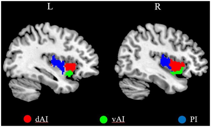
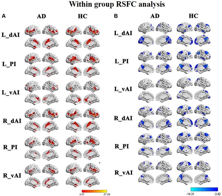
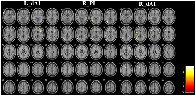
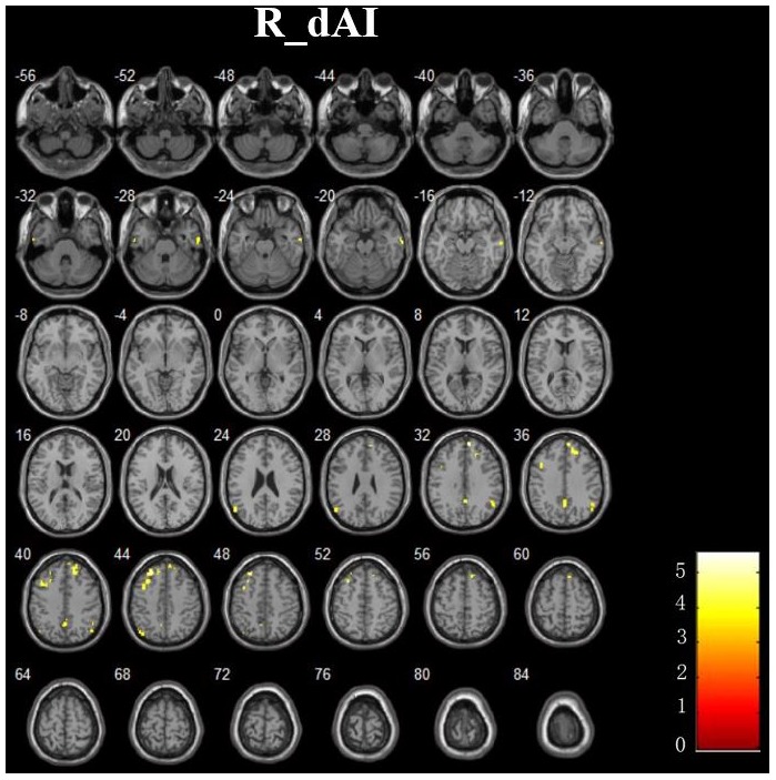
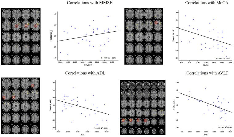
Recent researches have demonstrated that the insula is the crucial hub of the human brain networks and most vulnerable region of Alzheimer's disease (AD). However, little is known about the changes of functional connectivity of insular subregions in the AD patients. In this study, we collected resting-state functional magnetic resonance imaging (fMRI) data including 32 AD patients and 38 healthy controls (HCs). By defining three subregions of insula, we mapped whole-brain resting-state functional connectivity (RSFC) and identified several distinct RSFC patterns of the insular subregions: For positive connectivity, three cognitive-related RSFC patterns were identified within insula that suggest anterior-to-posterior functional subdivisions: (1) an dorsal anterior zone of the insula that exhibits RSFC with executive control network (ECN); (2) a ventral anterior zone of insula, exhibits functional connectivity with the salience network (SN); (3) a posterior zone along the insula exhibits functional connectivity with the sensorimotor network (SMN). In addition, we found significant negative connectivities between the each insular subregion and several special default mode network (DMN) regions. Compared with controls, the AD patients demonstrated distinct disruption of positive RSFCs in the different network (ECN and SMN), suggesting the impairment of the functional integrity. There were no differences of the positive RSFCs in the SN between the two groups. On the other hand, several DMN regions showed increased negative RSFCs to the sub-region of insula in the AD patients, indicating compensatory plasticity. Furthermore, these abnormal insular subregions RSFCs are closely correlated with cognitive performances in the AD patients. Our findings suggested that different insular subregions presented distinct RSFC patterns with various functional networks, which are differently affected in the AD patients.
Keywords: Alzheimer’s disease; fMRI; functional connectivity; insula; network.
Figures
Similar articles
-
Altered functional connectivity of insular subregions in subjective cognitive decline.Front Hum Neurosci. 2024 May 27;18:1404759. doi: 10.3389/fnhum.2024.1404759. eCollection 2024. Front Hum Neurosci. 2024. PMID: 38859994 Free PMC article.
-
Altered Functional Connectivity of Cognitive-Related Cerebellar Subregions in Alzheimer's Disease.Front Aging Neurosci. 2017 May 16;9:143. doi: 10.3389/fnagi.2017.00143. eCollection 2017. Front Aging Neurosci. 2017. PMID: 28559843 Free PMC article.
-
Abnormal Resting-State Functional Connectivity of Insular Subregions and Disrupted Correlation with Working Memory in Adults with Attention Deficit/Hyperactivity Disorder.Front Psychiatry. 2017 Oct 11;8:200. doi: 10.3389/fpsyt.2017.00200. eCollection 2017. Front Psychiatry. 2017. PMID: 29075206 Free PMC article.
-
Anterior insula as a gatekeeper of executive control.Neurosci Biobehav Rev. 2022 Aug;139:104736. doi: 10.1016/j.neubiorev.2022.104736. Epub 2022 Jun 11. Neurosci Biobehav Rev. 2022. PMID: 35700753 Review.
-
The Insula: A Stimulating Island of the Brain.Brain Sci. 2021 Nov 19;11(11):1533. doi: 10.3390/brainsci11111533. Brain Sci. 2021. PMID: 34827532 Free PMC article. Review.
Cited by
-
Abnormal static and dynamic functional connectivity of resting-state fMRI in multiple system atrophy.Aging (Albany NY). 2020 Aug 27;12(16):16341-16356. doi: 10.18632/aging.103676. Epub 2020 Aug 27. Aging (Albany NY). 2020. PMID: 32855356 Free PMC article.
-
Distinct resting-state functional connectivity patterns of Anterior Insula affected by smoking in mild cognitive impairment.Brain Imaging Behav. 2023 Aug;17(4):386-394. doi: 10.1007/s11682-023-00766-6. Epub 2023 May 27. Brain Imaging Behav. 2023. PMID: 37243752 Free PMC article.
-
Functional anomaly mapping reveals local and distant dysfunction caused by brain lesions.Neuroimage. 2020 Jul 15;215:116806. doi: 10.1016/j.neuroimage.2020.116806. Epub 2020 Apr 10. Neuroimage. 2020. PMID: 32278896 Free PMC article.
-
The abnormal accumulation of pathological proteins and compensatory functional connectivity enhancement of insula subdivisions in mild cognitive impairment.Front Aging Neurosci. 2025 Mar 18;17:1506478. doi: 10.3389/fnagi.2025.1506478. eCollection 2025. Front Aging Neurosci. 2025. PMID: 40171383 Free PMC article.
-
ACOEC-FD: Ant Colony Optimization for Learning Brain Effective Connectivity Networks From Functional MRI and Diffusion Tensor Imaging.Front Neurosci. 2019 Dec 12;13:1290. doi: 10.3389/fnins.2019.01290. eCollection 2019. Front Neurosci. 2019. PMID: 31920476 Free PMC article.
References
LinkOut - more resources
Full Text Sources
Other Literature Sources