

Review
doi: 10.3389/fmolb.2018.00031.
eCollection 2018.
A Glimpse Into the Structure and Function of Atypical Type I Chaperonins
Affiliations
- PMID: 29696145
- PMCID: PMC5904260
- DOI: 10.3389/fmolb.2018.00031
Item in Clipboard
Review
A Glimpse Into the Structure and Function of Atypical Type I Chaperonins
Front Mol Biosci.
.
Abstract
Chaperonins are a subclass of molecular chaperones that assist cellular proteins to fold and assemble into their native shape. Much work has been done on Type I chaperonins, which has elucidated their elegant mechanism. Some debate remains about the details in these mechanisms, but nonetheless the roles of these in helping protein folding have been understood in great depth. In this review we discuss the known functions of atypical Type I chaperonins, highlighting evolutionary aspects that might lead chaperonins to perform alternate functions.
Keywords: GroEL; GroES; Mycobacterium tuberculosis; Type I chaperonins; gene duplication; protein folding.
Figures
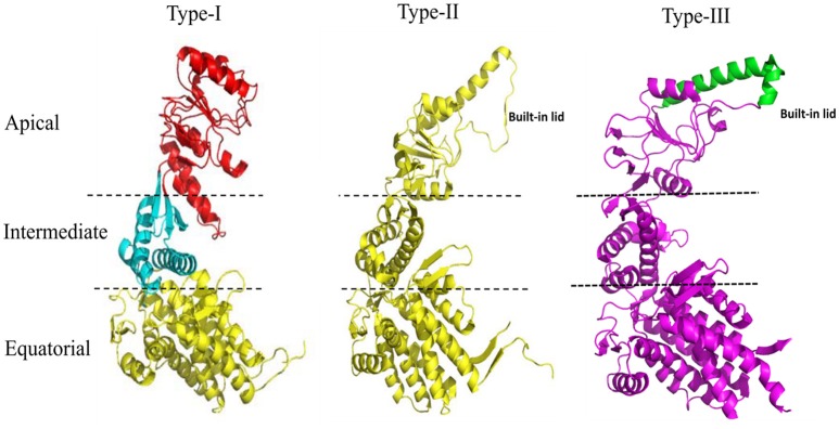
Structural features of the Type I, Type II, and Type III chaperonins. The comparative structure analysis of Type I, Type II, and Type III chaperonins. Structures were downloaded from the RCSB with codes of PDB: 1AON, 3RUW, and 5X9U, respectively. Type I chaperonin is demarcated into Apical, Intermediate, and Equatorial domains, analogous regions of which are shown in Type II and III chaperonins using dotted lines. Type II chaperonin has a characteristic built-in lid in the structure that plays the role of co-chaperonin GroES of Type I chaperonin. Type III chaperonins are structurally similar to Type II chaperonins in having built-in-lid. However, the sequence, structure and function of the lid are distinct in Type II and Type III chaperonins (An et al., 2017). The PyMOL program (PyMOL Molecular Graphics System, version 1.3) was used to generate this figure.
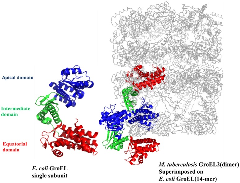
The crystal structure of M. tuberculosis GroEL2 superimposed on E. coli GroEL-ES structure. The structure of M. tuberculosis GroEL2 (PDB ID:1SJP) shows lower oligomeric status (dimer). Colored in blue, green, and red are the Apical, Intermediate and Equatorial domain, respectively. Compared to E. coli GroEL (PDB ID: 1AON) shown in gray color, the inter-subunit interaction is mediated through Apical domain in M. tuberculosis GroEL2 structure whereas inter-subunit interaction is through Equatorial domain in E. coli GroEL. Single subunit of M. tuberculosis GroEL2 is aligned to E. coli GroES bound GroEL ring representing asymmetric model. GroES structure has been removed for simplicity. A single subunit of E. coli GroEL has been shown with the same color-coded domains compared to M. tuberculosis GroEL2 for comparative analysis. The PyMOL program (PyMOL Molecular Graphics System, version 1.3) was used to generate this figure.
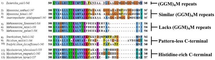
Multiple chaperonins in bacteria displaying diversity at C-terminal. Sequence alignment highlighting C-terminal regions of the representative bacterial GroEL homologs with the E. coli GroEL. The last C-terminal residues of selected multiple GroELs in different bacteria show divergence from the canonical (GGM)4M motif of the E. coli GroEL shown in dotted red box. Sequences were retrieved from www.uniprot.org and aligned in MEGA6 using MUSCLE algorithm (www.megasoftware.net ). Formatting of aligned sequences were done in Jalview alignment viewer (www.jalview.org ). Residues in the alignment follow the default Clustal color scheme of Jalview.
Similar articles
-
The unusual chaperonins of Mycobacterium tuberculosis.Tuberculosis (Edinb). 2005 Sep-Nov;85(5-6):385-94. doi: 10.1016/j.tube.2005.08.014. Epub 2005 Oct 25. Tuberculosis (Edinb). 2005. PMID: 16253564 Review.
-
Chaperonin-co-chaperonin interactions.Subcell Biochem. 2015;78:153-78. doi: 10.1007/978-3-319-11731-7_8. Subcell Biochem. 2015. PMID: 25487021 Review.
-
Chaperonins.Biochem J. 1998 Jul 15;333 ( Pt 2)(Pt 2):233-42. doi: 10.1042/bj3330233. Biochem J. 1998. PMID: 9657960 Free PMC article. Review.
-
Mycobacterial chaperonins: the tail wags the dog.FEMS Microbiol Lett. 2014 Jan;350(1):20-4. doi: 10.1111/1574-6968.12276. Epub 2013 Oct 7. FEMS Microbiol Lett. 2014. PMID: 24102684 Review.
-
The evolution of protein moonlighting: adaptive traps and promiscuity in the chaperonins.Biochem Soc Trans. 2014 Dec;42(6):1709-14. doi: 10.1042/BST20140225. Biochem Soc Trans. 2014. PMID: 25399594 Review.
Cited by
-
Chaperone-Mediated Autophagy and Its Implications for Neurodegeneration and Cancer.Int J Gen Med. 2022 Jun 15;15:5635-5649. doi: 10.2147/IJGM.S368364. eCollection 2022. Int J Gen Med. 2022. PMID: 35734200 Free PMC article. Review.
-
Implications of sperm heat shock protein 70-2 in bull fertility.Vet World. 2022 Jun;15(6):1456-1466. doi: 10.14202/vetworld.2022.1456-1466. Epub 2022 Jun 13. Vet World. 2022. PMID: 35993069 Free PMC article. Review.
-
Molecular, structural, and functional characterization of delta subunit of T-complex protein-1 from Leishmania donovani.Infect Immun. 2024 Oct 15;92(10):e0023424. doi: 10.1128/iai.00234-24. Epub 2024 Sep 9. Infect Immun. 2024. PMID: 39248465 Free PMC article.
-
Heat shock protein 60 and cardiovascular diseases: An intricate love-hate story.Med Res Rev. 2021 Jan;41(1):29-71. doi: 10.1002/med.21723. Epub 2020 Aug 17. Med Res Rev. 2021. PMID: 32808366 Free PMC article. Review.
-
The Role of Heat Shock Protein (Hsp) Chaperones in Environmental Stress Adaptation and Virulence of Plant Pathogenic Bacteria.Int J Mol Sci. 2025 Jan 9;26(2):528. doi: 10.3390/ijms26020528. Int J Mol Sci. 2025. PMID: 39859244 Free PMC article. Review.
References
Publication types
LinkOut - more resources
Full Text Sources
Other Literature Sources
Research Materials