Review
. 2018 Jun;11(2):272-279.
doi: 10.1007/s12178-018-9483-2.

Femoral Derotational Osteotomies

Affiliations
Review

Femoral Derotational Osteotomies

Manfred Nelitz. Curr Rev Musculoskelet Med. 2018 Jun.

Abstract

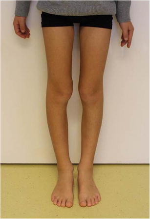
Purpose of review: Femoral derotational osteotomies are performed to correct residual symptomatic increased femoral torsion in adolescents and adults. Typical indications are anterior knee pain caused by patellar maltracking and patellofemoral instability. There is still no consensus as to what the correct indication is and which surgical techniques lead to the best outcomes in performing a femoral derotational osteotomy.

Recent findings: Good early clinical outcomes have been reported. However, long-term studies and data on return to play are lacking. Surgery often is performed according to the surgeon's experience. There is no evidence to support decisions regarding surgical technique or level of osteotomy. Femoral derotational osteotomy is the treatment of choice in patients with symptomatic excessive anteversion and torsional malalignment of the femur. Multiple techniques have shown good clinical results with high patient satisfaction. Future studies however must focus on radiographic and clinical assessment to understand different subtypes of torsional deformity and its implication on operative therapy.

Keywords: Femoral anteversion; Femoral derotational osteotomy; Hip pain; Intoeing; Knee pain; Patellofemoral instability.


Conflict of interest statement

Conflict of interest

Manfred Nelitz declares that he has no conflict of interest.

Human and animal rights and informed consent

This article does not contain any studies with human or animal subjects performed by any of the authors.

Figures

Fig. 1
Fig. 1
According to the technique described by Ruwe et al. [15], femoral anteversion can be measured clinically with the patient prone and knee flexed to 90°. The examiner feels for the greater trochanter of the femur and rotates the hip until the trochanter is most prominent laterally. In this position, the degree of femoral anteversion is estimated as the angle between the long axis of the tibia and a vertical line
Fig. 2
Fig. 2
View from the front visualizing the squinting patellae due to increased femoral anteversion
Fig. 3
Fig. 3
Radiographic measurement of femoral anteversion according to Rippstein [20] on an anteroposterior (AP) view of the hip with the patient supine and with the hips and knees flexed at 90°, the legs abducted 20°. Anteversion is measured as the angle between the horizontal axis and the axis of the femoral neck (red lines). Reprinted with permission
Fig. 4
Fig. 4
In the technique described by Waidelich et al. [16], the center of the femoral head on one transverse slice is connected to the center of an ellipse around the greater trochanter on another transverse slice. The axis in the distal part of the femur is a tangent to the posterior condyles on a transverse image. Femoral torsion is assessed by the angle between axes in the proximal and distal parts of the femur. In this case, the femoral torsion added up to 42°
Fig. 5
Fig. 5
Typical techniques of femoral derotational osteotomies. Proximal intertrochanteric osteotomy secured with an angle blade plate, diaphyseal osteotomies secured with an intramedullary rod, and distal supracondylar osteotomy secured with a locking plate
Fig. 6
Fig. 6
Intraoperative photograph demonstrating the two Schanz screws inserted into the femur proximal and distal to the planned osteotomy in order to facilitate derotation. The osteotomy was performed with drill holes and with an oscillating saw (a). Fixation with a locking plate after a femoral derotation osteotomy. The degree of the desired correction is visualized by the small vertical cuts (black arrows) (b)

References

    1. Kling TF, Jr, Hensinger RN. Angular and torsional deformities of the lower limbs in children. Clin Orthop Relat Res. 1983;176:136–147. - PubMed
    1. Fabry G, MacEwen GD, Shands AR. Torsion of the femur. A follow-up study in normal and abnormal conditions. J Bone Joint Surg Am. 1973;55:1726–1738. doi: 10.2106/00004623-197355080-00017. - DOI - PubMed
    1. Bruce WD, Stevens PM. Surgical correction of miserable malalignment syndrome. J Pediatr Orthop. 2004;24:392–396. doi: 10.1097/01241398-200407000-00009. - DOI - PubMed
    1. Parikh S, Noyes FR. Patellofemoral disorders: role of computed tomography and magnetic resonance imaging in defining abnormal rotational lower limb alignment. Sports Health. 2011;3:158–169. doi: 10.1177/1941738111399372. - DOI - PMC - PubMed
    1. Collado H, Fredericson M. Patellofemoral pain syndrome. Clin Sports Med. 2010;29:379–398. doi: 10.1016/j.csm.2010.03.012. - DOI - PubMed

LinkOut - more resources