

High-resolution computerized tomography changes in diffuse parenchymal lung disease from chronic hypersensitivity pneumonitis related to bird antigen
- PMID: 29697078
- PMCID: PMC5946554
- DOI: 10.4103/lungindia.lungindia_293_17
High-resolution computerized tomography changes in diffuse parenchymal lung disease from chronic hypersensitivity pneumonitis related to bird antigen
Abstract
Background: Chronic hypersensitivity pneumonitis (HP) is the most common cause of diffuse parenchymal lung disease (DPLD) in India. There is no data regarding the avian antigen exposure-associated DPLD from the country.
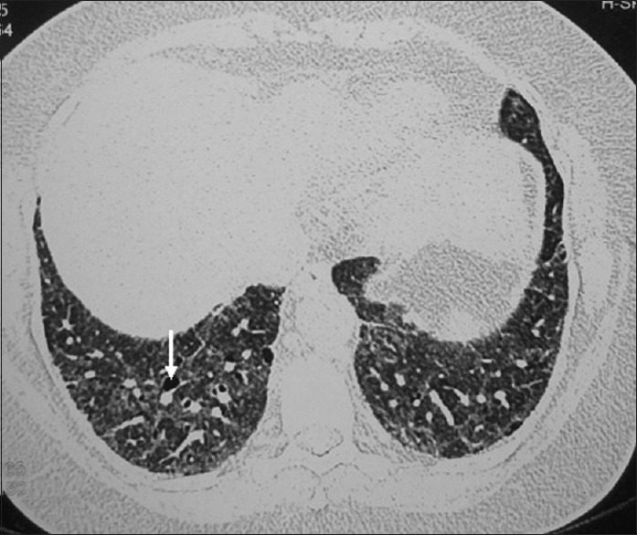
Methods: Chronic HP from exposure to avian antigen was diagnosed when the high resolution computerized tomography (HRCT) showed features for HP and was supported by the history of exposure to pigeons, the presence of precipitin antibodies (IgG) to avian antigen in high titre with negative rheumatoid factor, antinuclear antibody, and no clinical clue for a collagen vascular disease. The HRCT changes were noted on Likert scale (0-5) in terms of affection of peripheral and/or axial involvement, reticulation, honeycombing, haze, mosaic, traction bronchiectasis, pleural reactions, features of pulmonary hypertension, and air cysts. Cardiomegaly and independent cardiac chamber enlargement were also recorded.
Results: The lower lobes were predominantly (65.6%) affected with similar frequency (78.1) of peripheral and axial parenchymal affection. The parenchymal changes in HRCT were haze or ground-glass opacity (100%), mosaic appearance (93.75%), reticulations (68.75%), traction bronchiectasis (34.3%), air cysts (21.8%), and honeycombing (9.37%). Pleural reactions, though not described so far, were found in 50% of cases. Features of pulmonary hypertension (87.5%), cardiomegaly (50%), left and right atrial enlargement (81.2% and 78.1%), and right ventricular enlargement (31.2%) were the common echocardiography findings.
Conclusion: Chronic HP from avian exposure shows predominantly lower lobe involvement with haze, reticulation, features of pulmonary hypertension, and pleural reactions as common HRCT findings. The likelihood of pulmonary hypertension appears high and although honeycombing is often present, the classical UIP pattern has not been found.
Keywords: Diffuse parenchymal lung disease; forced vital capacity; high-resolution computerized tomography; hypersensitivity pneumonitis.
Conflict of interest statement
There are no conflicts of interest
Figures


Similar articles
-
Hypersensitivity Pneumonitis High-resolution Computed Tomography Findings, and Their Correlation with the Etiology and the Disease Duration.Prague Med Rep. 2020;121(3):133-141. doi: 10.14712/23362936.2020.12. Prague Med Rep. 2020. PMID: 33030142
-
Chronic hypersensitivity pneumonitis: high resolution computed tomography patterns and pulmonary function indices as prognostic determinants.Eur Radiol. 2012 Aug;22(8):1672-9. doi: 10.1007/s00330-012-2427-0. Epub 2012 Apr 1. Eur Radiol. 2012. PMID: 22466512
-
Compatible with fibrotic hypersensitivity pneumonitis on high-resolution computed tomography: from the ATS/JRS/ALAT 2020 hypersensitivity pneumonitis guidelines.J Thorac Dis. 2024 Apr 30;16(4):2353-2364. doi: 10.21037/jtd-23-1845. Epub 2024 Apr 12. J Thorac Dis. 2024. PMID: 38738228 Free PMC article.
-
Chronic hypersensitivity pneumonitis and pulmonary sarcoidosis: differentiation from usual interstitial pneumonia using high-resolution computed tomography.Semin Ultrasound CT MR. 2014 Feb;35(1):47-58. doi: 10.1053/j.sult.2013.10.006. Epub 2013 Oct 17. Semin Ultrasound CT MR. 2014. PMID: 24480143 Review.
-
CT signs and patterns of lung disease.Radiol Clin North Am. 2001 Nov;39(6):1115-35. doi: 10.1016/s0033-8389(05)70334-1. Radiol Clin North Am. 2001. PMID: 11699664 Review.
Cited by
-
Does avian antigen-induced chronic hypersensitivity pneumonitis lead to more severe PH than other causes of the same?Lung India. 2023 Mar-Apr;40(2):183-185. doi: 10.4103/lungindia.lungindia_323_22. Lung India. 2023. PMID: 37006108 Free PMC article. No abstract available.
References
-
- Malo JL, Zeiss CR. Occupational hypersensitivity pneumonitis after exposure to diphenylmethane diisocyanate. Am Rev Respir Dis. 1982;125:113–6. - PubMed
-
- Baur X. Hypersensitivity pneumonitis (extrinsic allergic alveolitis) induced by isocyanates. J Allergy Clin Immunol. 1995;95:1004–10. - PubMed
-
- Richerson HB, Bernstein IL, Fink JN, Hunninghake GW, Novey HS, Reed CE, et al. Guidelines for the clinical evaluation of hypersensitivity pneumonitis. Report of the subcommittee on hypersensitivity pneumonitis. J Allergy Clin Immunol. 1989;84:839–44. - PubMed
-
- Costabel U, Bonella F, Guzman J. Chronic hypersensitivity pneumonitis. Clin Chest Med. 2012;33:151–63. - PubMed
-
- Lacasse Y, Selman M, Costabel U, Dalphin JC, Ando M, Morell F, et al. Clinical diagnosis of hypersensitivity pneumonitis. Am J Respir Crit Care Med. 2003;168:952–8. - PubMed
LinkOut - more resources
Full Text Sources
Other Literature Sources
Research Materials
Miscellaneous
