. 2018 Aug;159(8):1641-1651.
doi: 10.1097/j.pain.0000000000001255.

Mice lacking Kcns1 in peripheral neurons show increased basal and neuropathic pain sensitivity

Affiliations

Mice lacking Kcns1 in peripheral neurons show increased basal and neuropathic pain sensitivity

Christoforos Tsantoulas et al. Pain. 2018 Aug.

Abstract

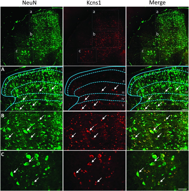
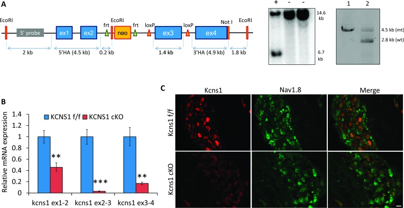
Voltage-gated potassium (Kv) channels are increasingly recognised as key regulators of nociceptive excitability. Kcns1 is one of the first potassium channels to be associated with neuronal hyperexcitability and mechanical sensitivity in the rat, as well as pain intensity and risk of developing chronic pain in humans. Here, we show that in mice, Kcns1 is predominantly expressed in the cell body and axons of myelinated sensory neurons positive for neurofilament-200, including Aδ-fiber nociceptors and low-threshold Aβ mechanoreceptors. In the spinal cord, Kcns1 was detected in laminae III to V of the dorsal horn where most sensory A fibers terminate, as well as large motoneurons of the ventral horn. To investigate Kcns1 function specifically in the periphery, we generated transgenic mice in which the gene is deleted in all sensory neurons but retained in the central nervous system. Kcns1 ablation resulted in a modest increase in basal mechanical pain, with no change in thermal pain processing. After neuropathic injury, Kcns1 KO mice exhibited exaggerated mechanical pain responses and hypersensitivity to both noxious and innocuous cold, consistent with increased A-fiber activity. Interestingly, Kcns1 deletion also improved locomotor performance in the rotarod test, indicative of augmented proprioceptive signalling. Our results suggest that restoring Kcns1 function in the periphery may be of some use in ameliorating mechanical and cold pain in chronic states.


Conflict of interest statement

Sponsorships or competing interests that may be relevant to content are disclosed at the end of this article.

Figures

Figure 1.
Figure 1.
Kcns1 expression in the mouse spinal cord. In the lumbar spinal cord, Kcns1 signal was detected in deeper laminae of the dorsal horn, where A fibers terminate. Thus, very little Kcns1 staining was observed in laminae I to II, but many immunoreactive neurons could be seen in laminae III to V (arrows in magnified inset a) and deeper laminae V to VI (inset b). Kcns1 labelling was also evident in large motoneurons of the ventral horn (inset c). Kcns1 appeared restricted to neuronal cells, as indicated by colocalisation with NeuN and the absence of staining in the white matter. Observations from n = 4 mice. Scale bars = 100 μm.
Figure 2.
Figure 2.
Kcns1 expression in the peripheral nerve. Kcns1 was detected along myelinated nerve fibers of the sciatic nerve, indicated by colocalisation with β3tubulin (white arrows). The Kcns1-expressing fibers were also found to express NF200 and CGRP, in agreement with the documented Kcns1 expression profile in the DRG. Some of the Kcns1-IR appeared to be nonneuronal (yellow arrows). Observations from n = 4 mice. Scale bars = 40 μm. CGRP, calcitonin gene-related peptide; DRG, dorsal root ganglion; IR, immunoreactivity.
Figure 3.
Figure 3.
Generation of Adv-CRE inducible Kcns1 conditional KO (cKO) mice. (A) Floxed Kcns1 knock-in mice were generated using homologous recombination in ES cells and subsequent blastocyst injections. (A) Schematic of the targeting construct is displayed here. Successful clones were identified using Southern blotting (middle, +: mutant clone) and PCR (right, 1: original targeting plasmid and 2: successful ES cell clone). The neomycin cassette was removed using an flp enzyme, and the resulting Kcns1 mice were crossed with the AdvCreERT2 line to generate an inducible knock-out of the Kcns1 gene in sensory and sympathetic neurons. (B) After induction of gene knock-out through tamoxifen treatment, DRG from cKO mice and floxed controls were analysed for Kcns1 mRNA expression by qPCR. Using primers amplifying within the deleted region of exon 3 (primer pairs 2-3 and 3-4), we found 97% and 89% reductions of Kcns1 mRNA in cKO mice compared with floxed controls. Using primers that bind before the targeted exon (exons 1-2), we also observed a 54% reduction in Kcns1 transcripts, most likely indicating degradation of incompletely formed mRNAs (**P<0.01, ***P<0.001; n = 3/group, the Student t test). (C) Kcns1 transcript reduction in cKO mice was also reflected at the protein level, as revealed by Kcns1 immunoreactivity in the DRG after tamoxifen treatment. As expected, Kcns1 protein was detected in medium–large, predominantly Nav1.8-negative neurons of floxed controls but was virtually absent from cKO mice. Observations from n = 3 mice/group. Scale bar = 40 μm. DRG, dorsal root ganglion; ES, embryonic stem; qPCR, quantitative real-time PCR.
Figure 4.
Figure 4.
Kcns1 deletion from sensory neurons affects basal pain processing. (A) Sensitivity to mechanical stimulation of the hind paw in conditional KO (Kcns1 cKO) and littermate control (Kcns1 f/f) male (left) and female (right) mice. Before induction of gene deletion with tamoxifen (“uninduced”), there is no difference in responses between genotypes (P > 0.05, the Student t test). However, 10 days after induction, there is a ∼20% decrease in pain thresholds in cKO mice compared with preinduced baseline (*P<0.05, **P<0.01, ***P<0.001; n = 11 male mice and n = 12 female mice/group; 2-way repeated-measures ANOVA with Tukey) (see also Suppl. Fig. 6, available online at http://links.lww.com/PAIN/A575). (B) Responses to noxious heat were not affected by Kcns1 deletion, as assessed with the Hargreaves method 10 days post-tamoxifen (P > 0.05; n = 11 male mice/group; 2-way repeated-measures ANOVA). (C) Kcns1 cKO mice and floxed littermates exhibited similar responses to noxious cold stimulation when placed on a 0°C cold plate (P > 0.05; n = 12 female mice/group; the Student t test). (D) Kcns1 deletion from sensory neurons resulted in enhanced locomotor performance in the rotarod test (*P<0.05; n = 12 female mice/group; the Student t test). ANOVA, analysis of variance.
Figure 5.
Figure 5.
Peripheral Kcns1 deletion triggers exaggerated pain phenotypes after nerve injury. (A) After tamoxifen treatment, the mice were subjected to partial nerve ligation and pain phenotypes monitored thereafter. Nerve injury resulted in pain hypersensitivity in both genotypes lasting for 3 weeks following the insult (#P < 0.05 vs post-TAM). In addition, pain thresholds in the cKO group were 27% lower compared with the floxed group at day 10 (**P < 0.01) and remained significantly lower thereafter (n = 11 male mice/group; 2-way repeated-measures ANOVA with Tukey). BL, pretamoxifen baseline; post-TAM, post-tamoxifen; dpi, days post injury. (B) Nerve damage decreased heat pain thresholds (###P < 0.001 vs post-TAM), but there was no difference between genotypes (n = 11 male mice/group; 2-way repeated-measures ANOVA). BL, pretamoxifen baseline; post-TAM, post-tamoxifen; dpi, days post injury. (C) Kcns1 deletion also affected behavioural responses to innocuous and noxious cold temperatures at 10 days after injury, manifested as increased paw lifting during a cold temperature ramp from 20 to 0°C. Left, cKO mice showed more paw lifts on average (P < 0.05, n = 11 male mice/group; the Student t test) during the ramp. Right, distribution of responses (P < 0.05; the Kolmogorov–Smirnov test) demonstrating cold hypersensitivity of cKO mice, particularly around temperatures of 10°C (P < 0.01) and 0°C (P < 0.001, n = 11/group; 2-way repeated-measures ANOVA with Tukey). ANOVA, analysis of variance; cKO, conditional KO.
Figure 6.
Figure 6.
Kcns1 protein expression after nerve injury in cKO and control mice. (A) Kcns1 expression on the contralateral (uninjured) and ipsilateral (injured) DRG from cKO and floxed littermate mice, 21 days after nerve damage. Kcns1 IR is largely attenuated by nerve injury in control mice; however, some Kcns1-positive neurons are still visible (arrows, top row). By contrast, no signal could be observed in either DRG from cKO mice (bottom row). (B) Quantification of percentages of Kcns1-positive neurons (**P < 0.01; n = 3/group; 1-way ANOVA with Tukey) and signal intensity (P > 0.05, n = 3/group; the Student t test). Scale bar = 40 μm. (C) Downregulation of Kcns1 in injured DRG neurons. Top panels, a fraction of small and medium–large sensory neurons are labelled with the nerve injury marker CSF1 at 21 days post-Seltzer. These injured neurons show no Kcns1 immunoreactivity (white arrows), which is however retained in CSF1-negative neurons (yellow arrows). Bottom panels, CSF1-positive neurons (arrows) show strong nuclear immunoreactivity for the injury marker ATF3. Scale bars, 40 μm. ANOVA, analysis of variance; cKO, conditional KO; DRG, dorsal root ganglion; IR, immunoreactivity.

References

    1. Bennett DL, Vincent A. Autoimmune pain: an emerging concept. Neurology 2012;79:1080–1. - PubMed
    1. Bocksteins E, Raes AL, Van de Vijver G, Bruyns T, Van Bogaert PP, Snyders DJ. Kv2.1 and silent Kv subunits underlie the delayed rectifier K+ current in cultured small mouse DRG neurons. Am J Physiol Cell Physiol 2009;296:C1271–1278. - PMC - PubMed
    1. Brosius Lutz A, Chung WS, Sloan SA, Carson GA, Zhou L, Lovelett E, Posada S, Zuchero JB, Barres BA. Schwann cells use TAM receptor-mediated phagocytosis in addition to autophagy to clear myelin in a mouse model of nerve injury. Proc Natl Acad Sci U S A 2017;114:E8072–80. - PMC - PubMed
    1. Busserolles J, Tsantoulas C, Eschalier A, Lopez Garcia JA. Potassium channels in neuropathic pain: advances, challenges, and emerging ideas. PAIN 2016;157(suppl 1):S7–14. - PubMed
    1. Calvo M, Richards N, Schmid AB, Barroso A, Zhu L, Ivulic D, Zhu N, Anwandter P, Bhat MA, Court FA, McMahon SB, Bennett DL. Altered potassium channel distribution and composition in myelinated axons suppresses hyperexcitability following injury. Elife 2016;5:e12661. - PMC - PubMed

Substances