

OPTIMAL MANAGEMENT OF PIGMENT EPITHELIAL DETACHMENTS IN EYES WITH NEOVASCULAR AGE-RELATED MACULAR DEGENERATION
- PMID: 29697591
- PMCID: PMC6221406
- DOI: 10.1097/IAE.0000000000002195
OPTIMAL MANAGEMENT OF PIGMENT EPITHELIAL DETACHMENTS IN EYES WITH NEOVASCULAR AGE-RELATED MACULAR DEGENERATION
Abstract
Purpose: This review aimed to determine the optimal management of retinal pigment epithelial detachments (PEDs) in neovascular age-related macular degeneration (nAMD) based on review of available evidence in the literature.
Methods: A comprehensive literature review evaluates previous retrospective and prospective studies that assessed the treatment of PEDs in nAMD.
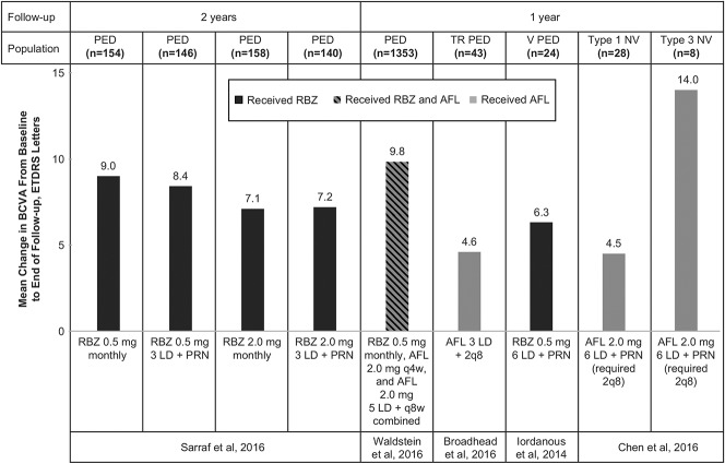
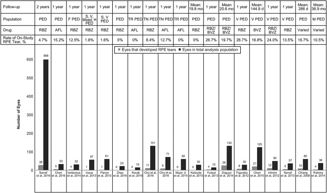
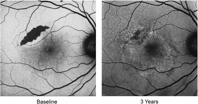
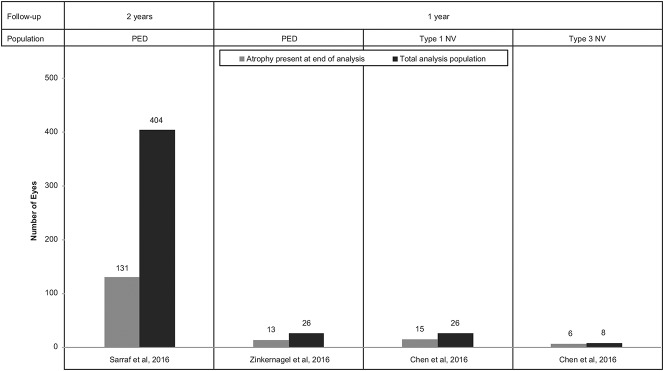
Results: Studies illustrated that anti-vascular endothelial growth factor (VEGF) therapy can be effective in eyes with PED secondary to nAMD. Similar visual outcomes are associated with different anti-VEGF treatments. Higher anti-VEGF doses may improve anatomical response, without correlation with vision improvement. Fibrovascular PEDs may be difficult to treat, but even these eyes can gain vision with anti-VEGF therapy. A retinal pigment epithelial tear may develop in 15% to 20% of eyes with PEDs after anti-VEGF therapy, especially in PEDs greater than 500 µm to 600 µm in height; however, vision may stabilize with continued therapy. Atrophy may complicate eyes with PED and nAMD after anti-VEGF therapy, especially in association with complete PED resolution.
Conclusion: Available literature suggests that anti-VEGF therapy is safe and efficacious for PED and nAMD. Treatment should focus on vision gains rather than PED resolution because there is no apparent correlation between anatomical and functional improvement in most eyes with PED and nAMD.
Figures
References
-
- Coscas F, Coscas G, Souied E, et al. Optical coherence tomography identification of occult choroidal neovascularization in age-related macular degeneration. Am J Ophthalmol 2007;144:592–599. - PubMed
-
- Schmidt-Erfurth U, Waldstein SM, Deak GG, et al. Pigment epithelial detachment followed by retinal cystoid degeneration leads to vision loss in treatment of neovascular age-related macular degeneration. Ophthalmology 2015;122:822–832. - PubMed
-
- Pauleikhoff D, Loffert D, Spital G, et al. Pigment epithelial detachment in the elderly. Clinical differentiation, natural course and pathogenetic implications. Graefes Arch Clin Exp Ophthalmol 2002;240:533–538. - PubMed
-
- Mariani A, Deli A, Ambresin A, Mantel I. Characteristics of eyes with secondary loss of visual acuity receiving variable dosing ranibizumab for neovascular age-related macular degeneration. Graefes Arch Clin Exp Ophthalmol 2011;249:1635–1642. - PubMed
Publication types
MeSH terms
Substances
LinkOut - more resources
Full Text Sources
Other Literature Sources
Medical
