. 2018 Jul;48(7):1199-1210.
doi: 10.1002/eji.201747442. Epub 2018 May 16.

Induced CNS expression of CXCL1 augments neurologic disease in a murine model of multiple sclerosis via enhanced neutrophil recruitment

Affiliations

Induced CNS expression of CXCL1 augments neurologic disease in a murine model of multiple sclerosis via enhanced neutrophil recruitment

Jonathan J Grist et al. Eur J Immunol. 2018 Jul.

Abstract

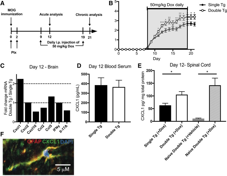
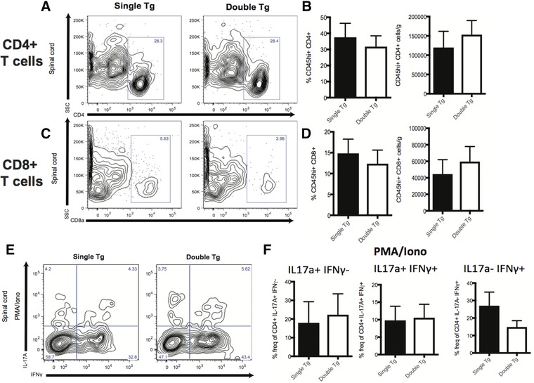
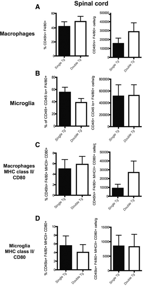
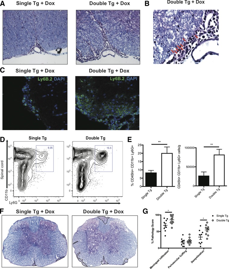
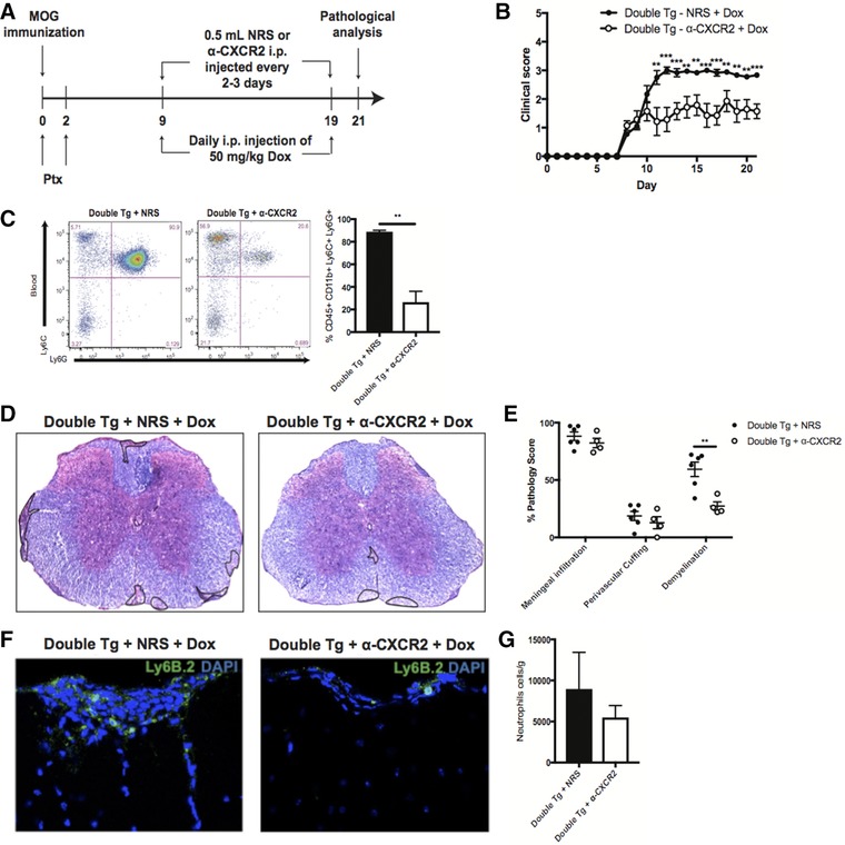
Increasing evidence points to an important role for neutrophils in participating in the pathogenesis of the human demyelinating disease MS and the animal model EAE. Therefore, a better understanding of the signals controlling migration of neutrophils as well as evaluating the role of these cells in demyelination is important to define cellular components that contribute to disease in MS patients. In this study, we examined the functional role of the chemokine CXCL1 in contributing to neuroinflammation and demyelination in EAE. Using transgenic mice in which expression of CXCL1 is under the control of a tetracycline-inducible promoter active within glial fibrillary acidic protein-positive cells, we have shown that sustained CXCL1 expression within the CNS increased the severity of clinical and histologic disease that was independent of an increase in the frequency of encephalitogenic Th1 and Th17 cells. Rather, disease was associated with enhanced recruitment of CD11b+ Ly6G+ neutrophils into the spinal cord. Targeting neutrophils resulted in a reduction in demyelination arguing for a role for these cells in myelin damage. Collectively, these findings emphasize that CXCL1-mediated attraction of neutrophils into the CNS augments demyelination suggesting that this signaling pathway may offer new targets for therapeutic intervention.

Keywords: Autoimmunity; Chemokines; Demyelination; Neuroinflammation; Neutrophils.


Figures

Figure 1
Figure 1
Dox‐induced CXCL1 expression increases clinical disease severity following MOG35–55‐induced EAE. (A) Schematic outline of experimental approach for disease induction and Dox administration. MOG, MOG35–55 peptide; PTX, pertussis toxin. (B) Clinical disease score was measured in MOG35–55‐immunized double tg mice (n  =  26) compared to single tg (n  =  29) following Dox treatment. Data were pooled from four independent experiments with between 6–8 mice per experiment. (C) mRNA transcripts of Cxcl1 and other pro‐inflammatory cytokines/chemokines in Dox‐treated double tg mice (n  =  2) compared to single tg mice (n  =  2) at day 12 p.i; data presented is representative of two independent experiments with a total of four mice per experimental group. CXCL1 levels in serum (D) and spinal cords (E) of MOG35–55‐immunized mice was determined by ELISA's at day 12 postimmunization. CXCL1 expression in spinal cords of either Dox‐ or vehicle‐treated double tg mice in the absence of peptide immunization (naïve) was also determined 3 days following last Dox injection (E); data is pooled from two independent experiments with between 2–4 mice for each experiment. (F) Representative immunofluorescence staining (60X) showing colocalization of CXCL1 protein (green) with GFAP‐positive (red) astrocytes in double tg mice at day 12 following Dox treatment (scale bar = 5 μm). Statistical analysis employed unpaired two‐tailed Student's t test; data presented as average ± SEM. *p < 0.05; **p < 0.01; ***p < 0.001.
Figure 2
Figure 2
T‐cell infiltration into the CNS is not altered following Dox‐induced expression of CXCL1. Spinal cords were removed at day 12 following MOG35–55 immunization of Dox‐treated double tg (n  =  10) and single tg (n  =  10) mice and the presence of CD45+CD4+ T cells (A, B) or CD45+CD8a+ T cells (C, D) determined by flow cytometry. The gating scheme employed is depicted in Supporting Information Fig. S1. Representative contour blots from experimental animals are shown in panels A and C. Data in panels B and D are pooled from two independent experiments with a minimum of three mice per experimental group; data is presented as mean ± SEM. (E and F) Expression of cytokines IL‐17A and/or IFN‐γ in Dox‐treated double tg or single tg following PMA/ionomycin treatment of CD4+ T‐cells isolated from spinal cords at day 12 following MOG35–55 immunization. Data in panel E are representative contour plots showing the results of intracellular staining for IL‐17A and IFN‐γ following PMA/ionomycin treatment of CD4+ T cells. The gating scheme employed is depicted in Supporting Information Fig. S2. Data in panel F represent quantification of intracellular cytokine staining and pooled from three independent experiments with a minimum of two mice per experimental group and presented as mean ± SEM and statistical analysis employed unpaired two‐tailed Student's t test.
Figure 3
Figure 3
Dox‐induced CXCL1 within the CNS does not increase microglia/macrophage activation. Dox‐treated double and single tg mice were sacrificed at day 12 following MOG35–55 immunization and spinal cords removed to assess macrophage (CD45hi F4/80+) (A) and microglia (CD45loF4/80+) (B) activation via flow cytometric staining between double tg (n  =  10) or single tg (n  =  10) mice. The gating scheme employed is depicted in Supporting Information Fig. S3. Surface expression of activation markers MHC class II and CD80 was also examined on macrophages (C) or microglia (D) between double tg (n  =  7) and single tg (n  =  7) mice. Data in panels A and B were pooled from three independent experiments; data in panels C and D from two independent experiments. Data are presented as mean ± SEM and statistical analysis employed unpaired two‐tailed Student's t test.
Figure 4
Figure 4
Neutrophil infiltration into the CNS is associated with increased demyelination. MOG35–55‐immunized double and single tg mice treated with Dox were sacrificed at day 12 postimmunization and spinal cords removed to assess histopathology and myeloid cell infiltration. (A) Representative H&E/Luxol fast blue staining of the spinal cords of experimental mice revealed increased inflammation within the anterior median fissure of double tg mice compared to single tg mice, 20X magnification. (B) Representative image of a spinal cord of Dox‐treated double tg mouse showing cells with multilobed nuclei (red arrows) characteristic of neutrophils, 60X magnification. (C) Representative immunofluorescent staining for the neutrophil‐specific surface antigen Ly6B.2 of double tg mice compared to single tg mice, 20X magnification. (D) Representative contour flow graphs showing increase in neutrophils (CD45hi CD11b+ Ly6G+) within the spinal cords of double tg mice compared to single tg mice. The gating scheme employed is depicted in Supporting Information Fig. S4. (E) Quantification of neutrophil flow staining in the spinal cords of double tg mice (n = 10) compared to single tg mice (n  =  10). Data in panel E were derived from three independent experiments and data presented as mean ± SEM. (F) Representative H&E/LFB stained spinal cord images (4X) demonstrated increased demyelination (outlined in black) in double tg mice compared to single tg mice at day 21 p.i. (G) Quantification of neuropathology indicated increased meningeal inflammation (p < 0.05), perivascular cuffing and demyelination (p < 0.05) at score at day 21 p.i. in double tg mice (n = 9) compared to single tg mice (n  =  9); data pooled from three independent experiments. Statistical analysis employed unpaired two‐tailed Student's t test, **p < 0.01.
Figure 5
Figure 5
Targeting neutrophils diminishes the severity of demyelination. MOG35–55‐immunized double and single tg mice treated with Dox were sacrificed at day 21 postimmunization and spinal cords removed to assess histopathology and myeloid cell infiltration. (A) Schematic outline for experimental design to target neutrophils in Dox‐treated double tg mice via injection of either anti‐CXCR2 or NRS control antibody. (B) Treatment with anti‐CXCR2 in Dox‐treated double tg mice (n  =  7) compared to animals treated with NRS (n  =  9); data pooled from three independent experiments with 2–3 mice per experiment. (C) Anti‐CXCR2 treatment in Dox‐treated double tg mice (n  =  3) resulted in reduced (p < 0.01) levels of neutrophils (CD45hi CD11b+ Ly6C+ Ly6G+) in blood compared to NRS‐treated mice (n  =  3); data derived from two independent experiments presented as mean ± SEM. The gating scheme employed is depicted in Supporting Information Fig. S5. (D) Representative H&E/LFB images depicting a reduction in demyelination (outlined in black) in Dox‐treated double tg compared to single tg mice and (E) quantification showed a significant (p < 0.01) reduction in demyelination in anti‐CXCR2 treated mice (n  =  6) compared to NRS‐treated mice (n  =  4); data is representative of two independent experiments. (F) Representative immunofluorescent staining for neutrophils (Ly6B.2) reveals fewer positive cells within the spinal cords of anti‐CXCR2‐treated mice compared to NRS‐treated mice. (G) Quantification of spinal cord Ly6B.2‐positive neutrophils reveals reduced numbers in anti‐CXCR2‐treated mice (n  =  3) compared to NRS‐treated mice (n  =  3); data derived from two independent experiments presented as mean ± SEM. **p < 0.01, ***p < 0.001.

Similar articles

Cited by

References

    1. Baker, D. and Amor, S. , Experimental autoimmune encephalomyelitis is a good model of multiple sclerosis if used wisely. Mult. Scler. Relat. Disord. 2014. 3: 555–564. - PubMed
    1. Martin, R. , McFarland, H. F. and McFarlin, D. E. , Immunological aspects of demyelinating diseases. Annu. Rev. Immunol. 1992. 10: 153–187. - PubMed
    1. Rao, P. and Segal, B. M. , Experimental autoimmune encephalomyelitis. Methods Mol. Biol. 2012. 900: 363–380. - PubMed
    1. Huseby, E. S. , Liggitt, D. , Brabb, T. , Schnabel, B. , Ohlen, C. and Goverman, J. , A pathogenic role for myelin‐specific CD8(+) T cells in a model for multiple sclerosis. J. Exp. Med. 2001. 194: 669–676. - PMC - PubMed
    1. Zamvil, S. , Nelson, P. , Trotter, J. , Mitchell, D. , Knobler, R. , Fritz, R. and Steinman, L. , T‐cell clones specific for myelin basic protein induce chronic relapsing paralysis and demyelination. Nature 1985. 317: 355–358. - PubMed

Publication types

MeSH terms