. 2018 Apr 26;13(4):e0196474.
doi: 10.1371/journal.pone.0196474. eCollection 2018.

Trypanosomal mitochondrial intermediate peptidase does not behave as a classical mitochondrial processing peptidase

Affiliations

Trypanosomal mitochondrial intermediate peptidase does not behave as a classical mitochondrial processing peptidase

Priscila Peña-Diaz et al. PLoS One. .

Abstract

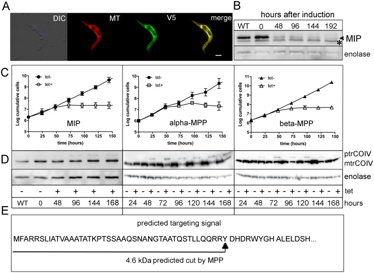
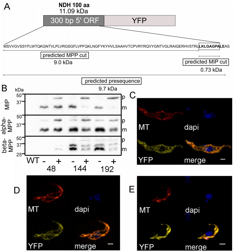
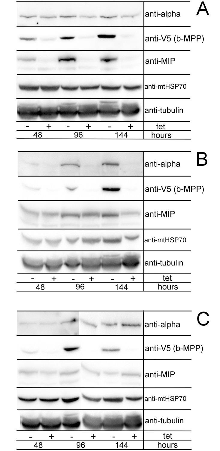
Upon their translocation into the mitochondrial matrix, the N-terminal pre-sequence of nuclear-encoded proteins undergoes cleavage by mitochondrial processing peptidases. Some proteins require more than a single processing step, which involves several peptidases. Down-regulation of the putative Trypanosoma brucei mitochondrial intermediate peptidase (MIP) homolog by RNAi renders the cells unable to grow after 48 hours of induction. Ablation of MIP results in the accumulation of the precursor of the trypanosomatid-specific trCOIV protein, the largest nuclear-encoded subunit of the cytochrome c oxidase complex in this flagellate. However, the trCOIV precursor of the same size accumulates also in trypanosomes in which either alpha or beta subunits of the mitochondrial processing peptidase (MPP) have been depleted. Using a chimeric protein that consists of the N-terminal sequence of a putative subunit of respiratory complex I fused to a yellow fluorescent protein, we assessed the accumulation of the precursor protein in trypanosomes, in which RNAi was induced against the alpha or beta subunits of MPP or MIP. The observed accumulation of precursors indicates MIP depletion affects the activity of the cannonical MPP, or at least one of its subunits.


Conflict of interest statement

Competing Interests: The authors have declared that no competing interests exist.

Figures

Fig 1
Fig 1. Phylogenetic analysis of T. brucei MIP and representative orthologs.
Maximum-likelihood tree of MIP was constructed using 50 taxa and 340 sites. Bootstrap support and posterior probability values were calculated for each branch using PhyML and MrBayes, respectively. Close relatives to MIP, thimet oligopeptidase homologs, were used as outgroups. Only bootstrap and posterior probability values greater than 50% and 0.5, respectively, are shown. Chosen taxa and accession numbers are shown in S2 Table.
Fig 2
Fig 2. Subcellular localization and depletion phenotype of MIP.
A) Immunofluorescence assay of MIP-V5 in procyclic stage T. brucei. Cells were incubated with MitoTracker (MT) (red), followed by immunodecoration with monoclonal anti-V5 antibody (V5) (green). DNA was stained using DAPI (blue). MIP was detected in the mitochondrion, as shown by the co-localization of the overexpressed protein with the MitoTracker signal (merge). In situ V5-tagged MIP showed identical localization results as the overexpressed protein (data not shown). B) Western blot analysis revealing the down-regulation of MIP by RNAi (indicated with a black arrowhead). The protein was efficiently depleted within 48 hrs of induction; * unspecific band. C) Growth curves of MIP, alpha-MPP and beta-MPP RNAi cell lines. Uninduced (black symbols) and RNAi-induced cell lines (white symbols). Three independent repeats were performed for each growth curve. D) Western blot analysis of trCOIV in cell lysates from RNAi cell lines for the respective peptidases, depicting accumulation of the precursor (ptrCOIV) and corresponding decrease of the mature protein (mtrCOIV). Enolase was used as a loading control. (+ tet) = RNAi-induced cells; (- tet) = uninduced cells. E) Schematic representation of the N-terminal sequence of trCOIV, displaying the predicted site for MPP cut. WT = wild-type cells; DIC = differential interference contrast; tet = tetracycline.
Fig 3
Fig 3. Processing of a YFP-chimera in the background of the mitochondrial processing peptidases.
Mitochondrial localization of the NDH-YFP chimera in trypanosomes RNAi-ablated for MIP. A) Schematic representation of the NDH-YFP chimera; the construct is composed of a stretch of 100 N-terminal amino acids from a putative subunit of complex I. The predicted sites for MPP and MIP cleavages are shown. B) Western blot analysis of the NDH-YFP protein in cells with inducible RNAi against the indicated peptidase. (+ tet), RNAi-induced cells; (- tet), uninduced cells; p, NDH-YFP precursor; m, mature NDH-YFP. Detection of the chimeric protein was perfomed using a polyclonal anti-GFP antibody. Subcellular distribution of this chimera was identical in RNAi cell lines of MIP (C), alpha-MPP (D) and beta-MPP (E). YFP signal is shown in yellow; MitoTracker (MT) is shown in red; DAPI stains DNA; merge shows all dyes at once. Scale bar is 1 μm.
Fig 4
Fig 4. Assessment of co-expression of MIP, alpha-MPP, and beta-MPP in an RNAi background of each of the three peptidases.
Beta-MPP was V5-tagged in situ in all three cell lines to monitor its expression by Western blot. 5 x 10⁶ cells/well were prepared in lysates for Western blot analysis. Samples were taken after 2, 4 and 6 days of RNAi induction. The levels of alpha-MPP, V5-tagged beta-MPP, MIP, and mtHSP70 were analyzed in all three cell lines. Tubulin was used as a loading control. A) MIP RNAi cell line. B) Alpha-MPP RNAi cell line. C) Beta-MPP RNAi cell line; (+ tet) = RNAi-induced cells; (- tet) = uninduced cells.

Similar articles

Cited by

References

    1. Neupert W. Protein import into mitochondria. Annu Rev Biochem. 1997;66: 863–917. doi: 10.1146/annurev.biochem.66.1.863 - DOI - PubMed
    1. Schmidt O, Pfanner N, Meisinger C. Mitochondrial protein import: from proteomics to functional mechanisms. Nat Rev Mol Cell Biol. 2010;11: 655–667. doi: 10.1038/nrm2959 - DOI - PubMed
    1. Hildenbeutel M, Habib SJ, Herrmann JM, Rapaport D. New insights into the mechanism of precursor protein insertion into the mitochondrial membranes. Int Rev Cell Mol Biol. 2008;268: 147–190. doi: 10.1016/S1937-6448(08)00805-8 - DOI - PubMed
    1. Eckers E, Cyrklaff M, Simpson L, Deponte M. Mitochondrial protein import pathways are functionally conserved among eukaryotes despite compositional diversity of the import machineries. Biol Chem. 2012;393: 513–524. doi: 10.1515/hsz-2011-0255 - DOI - PubMed
    1. Chacinska A, Koehler CM, Milenkovic D, Lithgow T, Pfanner N. Importing mitochondrial proteins: machineries and mechanisms. Cell. 2009;138: 628–644. doi: 10.1016/j.cell.2009.08.005 - DOI - PMC - PubMed

Publication types

MeSH terms

Substances

LinkOut - more resources