Profiling of epidermal lipids in a mouse model of dermatitis: Identification of potential biomarkers
- PMID: 29698466
- PMCID: PMC5919619
- DOI: 10.1371/journal.pone.0196595
Profiling of epidermal lipids in a mouse model of dermatitis: Identification of potential biomarkers
Abstract
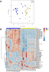
Lipids are important structural and functional components of the skin. Alterations in the lipid composition of the epidermis are associated with inflammation and can affect the barrier function of the skin. SHARPIN-deficient cpdm mice develop a chronic dermatitis with similarities to atopic dermatitis in humans. Here, we used a recently-developed approach named multiple reaction monitoring (MRM)-profiling and single ion monitoring to rapidly identify discriminative lipid ions. Shorter fatty acyl residues and increased relative amounts of sphingosine ceramides were observed in cpdm epidermis compared to wild type mice. These changes were accompanied by downregulation of the Fasn gene which encodes fatty acid synthase. A profile of diverse lipids was generated by fast screening of over 300 transitions (ion pairs). Tentative attribution of the most significant transitions was confirmed by product ion scan (MS/MS), and the MRM-profiling linear intensity response was validated with a C17-ceramide lipid standard. Relative quantification of sphingosine ceramides CerAS(d18:1/24:0)2OH, CerAS(d18:1/16:0)2OH and CerNS(d18:1/16:0) discriminated between the two groups with 100% accuracy, while the free fatty acids cerotic acid, 16-hydroxy palmitic acid, and docosahexaenoic acid (DHA) had 96.4% of accuracy. Validation by liquid chromatography tandem mass spectrometry (LC-MS/MS) of the above-mentioned ceramides was in agreement with MRM-profiling results. Identification and rapid monitoring of these lipids represent a tool to assess therapeutic outcomes in SHARPIN-deficient mice and other mouse models of dermatitis and may have diagnostic utility in atopic dermatitis.
Conflict of interest statement
Figures


Similar articles
-
Altered expression of epidermal lipid bio-synthesis enzymes in atopic dermatitis skin is accompanied by changes in stratum corneum lipid composition.J Dermatol Sci. 2017 Oct;88(1):57-66. doi: 10.1016/j.jdermsci.2017.05.005. Epub 2017 May 18. J Dermatol Sci. 2017. PMID: 28571749
-
Altered lipid properties of the stratum corneum in Canine Atopic Dermatitis.Biochim Biophys Acta Biomembr. 2018 Feb;1860(2):526-533. doi: 10.1016/j.bbamem.2017.11.013. Epub 2017 Nov 22. Biochim Biophys Acta Biomembr. 2018. PMID: 29175102
-
Lipid functions in skin: Differential effects of n-3 polyunsaturated fatty acids on cutaneous ceramides, in a human skin organ culture model.Biochim Biophys Acta Biomembr. 2017 Sep;1859(9 Pt B):1679-1689. doi: 10.1016/j.bbamem.2017.03.016. Epub 2017 Mar 21. Biochim Biophys Acta Biomembr. 2017. PMID: 28341437 Free PMC article.
-
Stratum Corneum Lipids: Their Role for the Skin Barrier Function in Healthy Subjects and Atopic Dermatitis Patients.Curr Probl Dermatol. 2016;49:8-26. doi: 10.1159/000441540. Epub 2016 Feb 4. Curr Probl Dermatol. 2016. PMID: 26844894 Review.
-
Epidermal Lipids: Key Mediators of Atopic Dermatitis Pathogenesis.Trends Mol Med. 2019 Jun;25(6):551-562. doi: 10.1016/j.molmed.2019.04.001. Epub 2019 May 1. Trends Mol Med. 2019. PMID: 31054869 Free PMC article. Review.
Cited by
-
Derailed Ceramide Metabolism in Atopic Dermatitis (AD): A Causal Starting Point for a Personalized (Basic) Therapy.Int J Mol Sci. 2019 Aug 15;20(16):3967. doi: 10.3390/ijms20163967. Int J Mol Sci. 2019. PMID: 31443157 Free PMC article. Review.
-
Suspect screening of exogenous compounds using multiple reaction screening (MRM) profiling in human urine samples.J Chromatogr B Analyt Technol Biomed Life Sci. 2022 Jun 30;1201-1202:123290. doi: 10.1016/j.jchromb.2022.123290. Epub 2022 May 12. J Chromatogr B Analyt Technol Biomed Life Sci. 2022. PMID: 35588643 Free PMC article.
-
Skeletal muscle undergoes fiber type metabolic switch without myosin heavy chain switch in response to defective fatty acid oxidation.Mol Metab. 2022 May;59:101456. doi: 10.1016/j.molmet.2022.101456. Epub 2022 Feb 9. Mol Metab. 2022. PMID: 35150906 Free PMC article.
-
An Integrative Proteomic/Lipidomic Analysis of the Gold Nanoparticle Biocorona in Healthy and Obese Conditions.Appl In Vitro Toxicol. 2019 Sep 1;5(3):150-166. doi: 10.1089/aivt.2019.0005. Epub 2019 Sep 17. Appl In Vitro Toxicol. 2019. PMID: 32292798 Free PMC article.
-
Octanoate is differentially metabolized in liver and muscle and fails to rescue cardiomyopathy in CPT2 deficiency.J Lipid Res. 2021;62:100069. doi: 10.1016/j.jlr.2021.100069. Epub 2021 Mar 20. J Lipid Res. 2021. PMID: 33757734 Free PMC article.
References
-
- Kendall AC, Nicolaou A. Bioactive lipid mediators in skin inflammation and immunity. Prog Lipid Res. 2013. January;52(1):141–64. doi: 10.1016/j.plipres.2012.10.003 - DOI - PubMed
-
- van Smeden J, Janssens M, Gooris GS, Bouwstra JA. The important role of stratum corneum lipids for the cutaneous barrier function. Biochim Biophys Acta—Mol Cell Biol Lipids. 2014. March;1841(3):295–313. - PubMed
-
- Flohr C, Mann J. New insights into the epidemiology of childhood atopic dermatitis. Allergy. 2014. January;69(1):3–16. doi: 10.1111/all.12270 - DOI - PubMed
-
- Maksimović N, Janković S, Marinković J, Sekulović LK, Zivković Z, Spirić VT. Health-related quality of life in patients with atopic dermatitis. J Dermatol. 2012. January;39(1):42–7. doi: 10.1111/j.1346-8138.2011.01295.x - DOI - PubMed
Publication types
MeSH terms
Substances
Grants and funding
LinkOut - more resources
Full Text Sources
Other Literature Sources
Molecular Biology Databases
Miscellaneous

