Comment
. 2018 Oct:73:180-191.
doi: 10.1016/j.bbi.2018.04.012. Epub 2018 Apr 24.

Distinct characteristics of hippocampal pathogenic TH17 cells in a mouse model of depression

Affiliations
Comment

Distinct characteristics of hippocampal pathogenic TH17 cells in a mouse model of depression

Eléonore Beurel et al. Brain Behav Immun. 2018 Oct.

Abstract

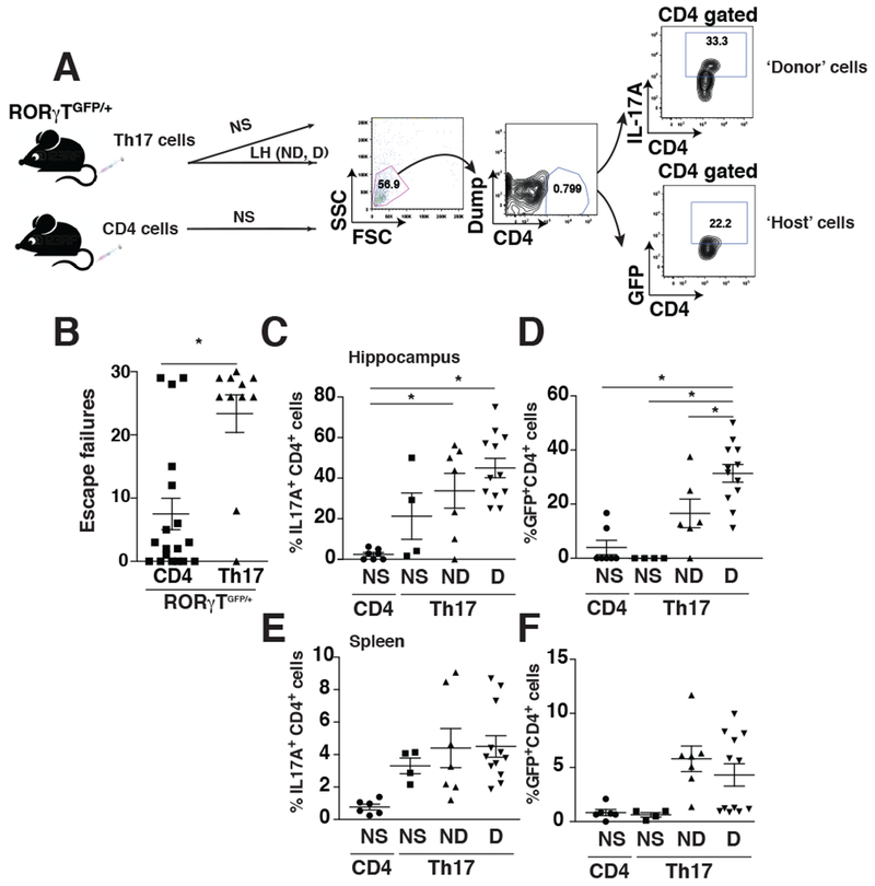
Increasing evidence indicates that multiple actions of the immune system are closely intertwined with the development of depression and subsequent recovery processes. One of these interactions is substantial evidence that the TH17 subtype of CD4+ T cells promotes susceptibility to depression-like behaviors in mice. Comparing subtypes of CD4+ T cells, we found that administration of TH17 cells, but not TH1 cells or TREGS, promoted susceptibility to learned-helplessness depressive-like behavior and accumulated in the hippocampus of learned helpless mice. Adoptively transferred TH17 cells into Rag2-/- mice that are devoid of endogenous T cells increased susceptibility to learned helplessness, demonstrating that increased peripheral TH17 cells are capable of modulating depression-like behavior. Moreover, in wild-type mice, adoptively transferred TH17 cells accumulated in the hippocampus of learned-helpless mice and induced endogenous TH17 cell differentiation. Hippocampal TH17 cells from learned-helpless mice expressed markers of pathogenic TH17 cells (CCR6, IL-23R) and of follicular cells (CXCR5, PD-1), indicating that the hippocampal cells are TFH-17-like cells. Knockout of CCR6 blocked TH17 cells from promoting learned helplessness, which was associated with increased expression of PD-1 in CCR6-deficient TH17 cells. In summary, these results reinforce the conclusion that depression-like behaviors are selectively facilitated by TH17 cells, and revealed that these cells in the hippocampus of learned helpless mice display characteristics of TFH17-like cells, which may contribute to their pathogenic actions in promoting depression.

Keywords: Depression; Th17.


Conflict of interest statement

Financial disclosures

The authors have no financial interests or conflicts of interest.

Figures

Figure 1:
Figure 1:
TH17 cells promote learned-helplessness and accumulate in the hippocampus and PFC. (A) CD4+ T cells were differentiated toward either TH17, TH1 or undifferentiated CD4+ cells for 4 days in vitro and adoptively transferred to male and female Rag2−/− (B) or male wild-type (C) mice. In (B) 18% of the CD4+ cells were TH17 cells in mice receiving TH17 cells, and 39% of the CD4+ cells were TH1 cells in mice receiving TH1 cells. In mice receiving CD4+ cells, less than 1% of CD4+ cells were TH17 cells or TH1 cells. 48 h after transfer mice were subjected to the reduced paradigm of learned-helplessness. The number of escape failures was recorded. Each symbol represents an individual mouse. Mean ± SEM, one-way ANOVA, F(2,36)=4.037, Bonferroni post hoc test *p<0.05 (n=7-14). (C) Mice were subjected or not to the learned-helplessness (LH) paradigm, and were separated into 3 groups: non-shocked (NS), shocked and exhibiting >15/30 escape failures (D), or shocked and not learned-helpless (<15 escape failures, ND). (D-F) Mice were perfused, and hippocampus and PFC were processed to obtain dissociated cells. Cells were stained for CD4 and IL-17A (D), IFNγ (E) or Foxp3 (F), and gated on CD4+ cells and analyzed by flow cytometry. Data represent mean ± SEM, one-way ANOVA, F(5, 30)=10.26 (E), F(5,39)=14.64 (F), Bonferroni post hoc test *p<0.05 (n=4-11).
Figure 2:
Figure 2:
Adoptively transferred TH17 cells accumulate in the hippocampus after the learned-helplessness paradigm. (A) CD45.2 wild-type recipient mice received donor CD45.1+CD4+ T cells or CD45.1+ TH17 cells, and 48 h later were subjected to the learned-helplessness paradigm. 86% of the CD4+T cells transferred in the TH17 cells preparation were TH17 cells, whereas less than 1% TH17 cells were detected in the CD4 preparation, and 99.3% of the injected CD4 cells were CD45.1. (B) CD4+ CD45.1+ (donor cells) or CD4+CD45.2+ (recipient cells) present in the hippocampus of mice adoptively transferred with CD4 cells or TH17 cells were analyzed by flow cytometry after gating on CD4+ cells (C) CD45.1+CD4+ (donor cells) or CD45.2+IL-17A+CD4+ (recipient cells) present in the hippocampus of mice adoptively transferred with CD4 cells or TH17 cells were analyzed by flow cytometry after gating on CD45+CD4+ cells. Each symbol represents an individual mouse. Data represent mean±SEM, one-way ANOVA, F(3, 16) = 9.522, Bonferroni post-hoc test *p<0.05 (n=5). (D) CD45.1+IL-17A+CD4+ (donor cells) or CD45.2+IL-17A+CD4+ (recipient cells) present in the hippocampus of mice adoptively transferred with CD4 cells or TH17 cells were analyzed by flow cytometry after gating on CD45+IL-17A+CD4+ cells. Each symbol represents an individual mouse. Data represent mean±SEM, one-way ANOVA, F(3, 14)=9,795, Bonferroni post-hoc test *p<0.05 (n=4-5).
Figure 3:
Figure 3:
Induction of learned-helplessness increases the induction of GFP in RORγTGFP/+ mice. (A-B) RORγTGFP/+ mice adoptively transferred with undifferentiated CD4 cells or TH17 cells 48 h before being subjected to the reduced learned-helplessness paradigm, and the number of escape failures was recorded. Each symbol represents an individual mouse. Data represent mean±SEM, Mann Whitney, U=37.5, *p<0.05, (n=11-18). (C-F) Unshocked (NS) mice or mice transferred with TH17 cells (Th17), or mice exhibiting learned-helplessness (D) or non-learned-helpless mice (ND) that received TH17 cells were perfused after the last shock and hippocampal (C-D) or splenic (E-F) cells were dissociated. CD4+ T cells were stained for IL-17A, and both IL-17A+CD4+ T cells (C, E) and GFP+CD4+ T cells (D, F) were evaluated by flow cytometry after gating on CD4+ cells as described in panel (A). Each symbol represents an individual mouse. Data represent mean ±SEM, one-way ANOVA, F(3, 26)= 9,492 (B), F(3,25)=15.75 (C), Bonferroni post hoc test *p<0.05 (n=4-12).
Figure 4:
Figure 4:
Follicular and pathogenic markers are elevated in the hippocampus of learned-helpless mice. (A) Mice were subjected or not to the learned-helplessness paradigm, and mice were separated into 3 groups: learned-helpless (D), non-learned-helpless (ND) and non-shocked (NS) as described in Fig 1D. Immediately after the last foot shocks, hippocampi were collected and mRNA extracted to measure by qRT-PCR: IL-17A mRNA (A), an array of TH17 cell-related genes (B), ICOS mRNA (C), c-maf mRNA (D), IL-27 mRNA (E) and Bcl6 mRNA (F). Genes upregulated or downregulated more than 4-fold are reported in (B). Each symbol represents an individual mouse. Data represent mean±SEM, One-way ANOVA, F(2, 12)=6.212 (A), F(2, 25) = 20.21 (C), F(2,18)=57.66 (D), Bonferroni post-hoc test *p<0.05 (n=4-10). (G-I) Mice were subjected to the learned-helplessness paradigm, or not, and mice were separated into 3 groups: unshocked (NS), learned-helpless (D) and non-learned-helpless (ND) mice. Immediately after the last foot shocks, mice were perfused and hippocampi were collected. Dissociated cells were stained for CD4, IL-17A, and either CCR6 (G) or IL-23R (I), and gated on IL-17A+ CD4+ cells. (H) mRNA was extracted from hippocampus and CCL20 expression was analyzed by qRT-PCR. Each symbol represents an individual mouse. Data represent mean±SEM, one-way ANOVA, F(2, 23)= 11.59 (A), F(2, 15)=16.50 (B), F(2,33)=8.034 (C), Bonferroni post-hoc test *p<0.05 (n=8-17).
Figure 5:
Figure 5:
TH17 cells associated with learned helplessness present a follicular phenotype. Mice were subjected to the learned-helplessness paradigm, or not, and mice were separated into 3 groups: unshocked (NS), learned-helpless (D) and non-learned-helpless (ND) as described in Fig 1D. Immediately after the last foot shocks, mice were perfused and hippocampi were collected. Dissociated cells were stained for CD4, CXCR5 and gated on CD4+ cells (A-B) or cells were stained for CD4, CXCR5, IL-17A and gated (C-D) or not (E-F) on CXCR5+CD4+ cells; or cells were stained for CD4, CXCR5 and either IFNγ (G-H), or ICOS (I-J), or IL-21 (M-N), and gated on CXCR5+CD4+ cells (G-L); or cells were stained for CD4, ICOS and gated on CD4+ cells (K-L) or CD4, IL-21 and gated on CD4+ICOS+ cells (O-P). Representative plots of flow cytometry are presented in A, C, E, G, I, K, M and O, and quantifications are presented in B, D, F, H, J, L, N and P. Each symbol represents an individual mouse. Data represent mean± SEM, one-way ANOVA, F(2,32)=13.33 (B), F(2,38)= 11.20 (D), Bonferroni post-hoc test and Mann-Whitney, U=14 (J), *p<0.05 (n=4-20).
Figure 6:
Figure 6:
TFH-like 17 cells have increased PD-1 levels and express CCR6 and IL-23R. Mice were subjected to the learned-helplessness paradigm, and were separated into 2 groups: learned-helpless (D) and non-learned-helpless (ND). Immediately after the last foot shocks, mice were perfused and hippocampi were collected. Dissociated cells were stained for CD4, CXCR5, IL-17A, and either PD-1 (A-B), or CCR7 (C-D), or BCL-6 (E-F), and gated on IL-17A+CD4+ cells (A-F). Each symbol represents an individual mouse. Data represent mean±SEM, t-test, t(10)=2.729 *p<0.05 (n=5-7). (G-N) Mice were subjected to the learned-helplessness paradigm, or not, and mice were separated into 3 groups: unshocked (NS), learned-helpless (D) and non-learned-helpless (ND) mice. Immediately after the last foot shocks, mice were perfused and hippocampi were collected. Dissociated cells were stained for CD4, CXCR5, IL-17A, and either CCR6 (G-H, K-L) or IL-23R (I-J, M-N), and gated on CXCR5+IL-17A+CD4+ cells (G-J) or on CXCR5+CD4+cells (K-M). Representative plots of flow cytometry are presented in G, I, K and M, and quantifications are presented in H, J, L and N. Each symbol represents an individual mouse. Data represent mean± SEM, Mann-Whitney, U=17 (H), U=20 (J), *p<0.05 (n=8-17).
Figure 7:
Figure 7:
TFH-like 17 cells originate from the host. CD45.2 wild-type recipient mice received donor CD45.1+CD4+ T cells or CD45.1+ TH17 cells, and 48 h later were subjected to the learned-helplessness paradigm. 86% of the CD4+T cells transferred in the TH17 cells group were TH17 cells, whereas less than 1% TH17 cells were detected in the CD4 group, and 99.3% of the CD4 injected cells were CD45.1 as described in Fig 2A. (A) CD4+CD45.1+ (donor cells) or CD4+CD45.2+ (recipient cells) present in the hippocampus of mice adoptively transferred with CD4 or TH17 cells were analyzed by flow cytometry after gating on CD4+ cells. CD45.1+CXCR5+IL-17A+CD4+ (donor cells) or CD45.2+CXCR5+IL-17A+CD4+ (recipient cells) present in the hippocampus (B) or spleen (C) of mice adoptively transferred with CD4 or TH17 cells were analyzed by flow cytometry after gating on CD45+CXCR5+IL-17A+CD4+ cells as described in panel (A). Each symbol represents an individual mouse. Data represent mean±SEM, one-way ANOVA, F(2,14)=22.9 (A), F(3,16)=34.35 (B), Bonferroni post-hoc test *p<0.05 (n=5).
Figure 8:
Figure 8:
CCR6 expression is required for TH17 cells to promote learned-helplessness. Male and female mice received TH17 cells transduced with either a scrambled siRNA (siSCR) or siRNA targeting CCR6 (siCCR6) and 48 h later were subjected to the reduced paradigm of learned-helplessness. (A) Before transfer, TH17 cells transduced with either a scrambled siRNA (47.4% CD4 are TH17 cells) or siRNA targeting CCR6 (43.5% CD4 are TH17 cells) were stained for CCR6, IL-17A and CD4 for analysis by flow cytometry. Mean ± SEM, Mann-Whitney, U=0, *p<0.05 (n=3-4). (B) The number of escape failures was recorded. Each symbol represents an individual mouse. Mean ± SEM, t-test, t(20)=2.132, *p<0.05 (n=11). Immediately after the last foot shocks, mice were perfused and hippocampi were collected. Dissociated cells were stained for IL-17A and CD4 (C), CD4 (C), Foxp3 and CD4 (E), F4/80 and CD45 (F), F4/80 and CD45 (G), CD11c and CD45 (H) and gated on CD4+ cells (C-E). Data represent mean±SEM, (n=5). (I) Mice received TH17 cells differentiated from wild-type CD4 cells (WT, 33% CD4 are TH17 cells) or CCR6-deficient CD4 cells (CCR6 KO, 28% CD4 are TH17 cells) and 48 h later were subjected to the reduced paradigm of learned-helplessness. The number of escape failures was recorded. Each symbol represents an individual mouse. Mean ± SEM, Mann-Whitney, *p<0.05 (n=10). Immediately after the last foot shocks, mice were perfused and hippocampi were collected. Dissociated cells were stained for IL-17A and CD4, and gated on CD4+ cells (J) or stained for PD-1, IL-17A, and CD4 and gated on IL-17A+ CD4+ cells (K). Data represent mean±SEM, Mann-Whitney U=21 (I) and Wilcoxon test, W=−19 (K), *p<0.05 (n=9).

Comment on

References

    1. Acosta-Rodriguez EV, Rivino L, Geginat J, Jarrossay D, Gattorno M, Lanzavecchia A, Sallusto F, and Napolitani G (2007). Surface phenotype and antigenic specificity of human interleukin 17-producing T helper memory cells. Nat Immunol 8, 639–646. - PubMed
    1. Annunziato F, Cosmi L, Santarlasci V, Maggi L, Liotta F, Mazzinghi B, Parente E, Fili L, Ferri S, Frosali F, et al. (2007). Phenotypic and functional features of human Th17 cells. J Exp Med 204, 1849–1861. - PMC - PubMed
    1. Awasthi A, and Kuchroo VK (2009). Th17 cells: from precursors to players in inflammation and infection. Int Immunol 21, 489–498. - PMC - PubMed
    1. Bauquet AT, Jin H, Paterson AM, Mitsdoerffer M, Ho IC, Sharpe AH, and Kuchroo VK (2009). The costimulatory molecule ICOS regulates the expression of c-Maf and IL-21 in the development of follicular T helper cells and TH-17 cells. Nat Immunol 10, 167–175. - PMC - PubMed
    1. Beurel E, Harrington LE, and Jope RS (2013). Inflammatory T helper 17 cells promote depression-like behavior in mice. Biol Psychiatry 73, 622–630. - PMC - PubMed

Publication types