Review
. 2011 Jul 2;10(4):239-249.
doi: 10.1007/s12522-011-0099-0. eCollection 2011 Dec.

Cellular and molecular mechanisms of various types of oocyte aging

Affiliations
Review

Cellular and molecular mechanisms of various types of oocyte aging

Toshifumi Takahashi et al. Reprod Med Biol. .

Abstract

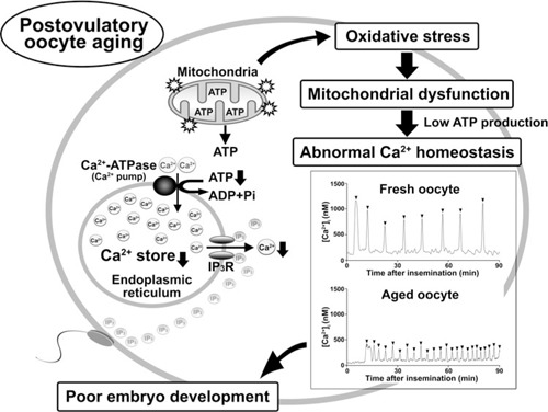
It is well established that age-related decline of a woman's fertility is related to the poor developmental potential of her gametes. The age-associated decline in female fertility is largely attributable to the oocyte aging caused by ovarian aging. Age-associated oocyte aging results in a decrease in oocyte quality. In contrast to ovarian aging, there is a concept of postovulatory oocyte aging. Postovulatory aging of oocytes, not being fertilized for a prolonged time after ovulation, is known to significantly affect the development of oocytes. Both categories of oocyte aging have similar phenotypes of reproductive failure. However, the mechanisms of the decline in oocyte quality are not necessarily equivalent. An age-dependent increase in aneuploidy is a key determinant of oocyte quality. The reduced expression of molecules regulating cell cycle control during meiosis might be involved in the age-dependent increase in aneuploidy. The mechanism of age-associated oocyte aging might be involved in mitochondrial dysfunction, whose etiologies are still unknown. Alternatively, the mechanism of postovulatory oocyte aging might be involved in reactive oxygen species-induced mitochondrial injury pathways followed by abnormal intracellular Ca2+ regulation of the endoplasmic reticulum. We suggest that future research into the mechanism of oocyte aging will be necessary to develop a method to rescue the poor developmental potential of aged oocytes.

Keywords: Calcium regulation; Oocyte aging; Ovarian aging; Oxidative stress; Postovulatory oocyte aging.


Conflict of interest statement

The authors have nothing to disclose.

Figures

Figure 1
Figure 1
Scheme of the mechanism of poor embryo development in postovulatory oocyte aging. We show the model of the mechanism of poor embryo development in postovulatory‐aged oocytes. Postovulatory aging of oocytes results in increase in mitochondrial oxidative stress. Oxidative stress‐induced mitochondrial dysfunction results in low ATP production followed by impairment of intracellular Ca2+ regulation, such as decrease in the Ca2+ stores of the endoplasmic reticulum (ER) and the Ca2+ release from the ER via inositol 1,4,5‐triphosphate (InsP3) receptor. When postovulatory aged‐oocytes, which are impaired in the intracellular Ca2+ regulations, are fertilized with sperm, the abnormal Ca2+ oscillations occur at fertilization. Representative data show that sperm triggers Ca2+ oscillations in the fresh (14 h after hCG treatment) and the aged (20 h after hCG treatment) mouse oocytes. The patterns of Ca2+ oscillations at fertilization are changed by postovulatory oocyte aging: In the aged oocytes, the abnormal Ca2+ oscillations with lower amplitude and high frequency are shown. The abnormal Ca2+ oscillations may result in poor embryo development in postovulatory‐aged oocytes. Arrowheads indicate the individual Ca2+ oscillations. IP3, InsP3. IP3R, InsP3 receptor

Similar articles

Cited by

References

    1. Balasch J. Ageing and infertility: an overview. Gynecol Endocrinol, 2010, 26, 855–860 10.3109/09513590.2010.501889 - DOI - PubMed
    1. Mouzon J, Lancaster P, Nygren KG, Sullivan E, Zegers‐Hochschild F, Mansour R, Ishihara O, Adamson D. World collaborative report on assisted reproductive technology, 2002. Hum Reprod, 2009, 24, 2310–2320 10.1093/humrep/dep098 - DOI - PubMed
    1. Baird DT, Collins J, Egozcue J, Evers LH, Gianaroli L, Leridon H, Sunde A, Templeton A, Steirteghem A, Cohen J, Crosignani PG, Devroey P et al. Fertility and ageing. Hum Reprod Updat, 2005, 11, 261–276 10.1093/humupd/dmi006 - DOI - PubMed
    1. Leridon H. Can assisted reproduction technology compensate for the natural decline in fertility with age? A model assessment. Hum Reprod, 2004, 19, 1548–1553 10.1093/humrep/deh304 - DOI - PubMed
    1. te Velde ER, Pearson PL. The variability of female reproductive ageing. Hum Reprod Updat, 2002, 8, 141–154 10.1093/humupd/8.2.141 - DOI - PubMed