Review
. 2008 Apr 17;7(2):91-103.
doi: 10.1111/j.1447-0578.2008.00205.x. eCollection 2008 Jun.

Angiogenesis in the human corpus luteum

Affiliations
Review

Angiogenesis in the human corpus luteum

Norihiro Sugino et al. Reprod Med Biol. .

Abstract

Angiogenesis is important for the formation and development of the corpus luteum and for maintenance of luteal function. Blood vessel regression is an important physiological phenomenon in the corpus luteum, which is associated with tissue involution during structural luteolysis. Angiogenesis actively occurs during the early luteal phase and is completed by the mid-luteal phase. Perivascular cells (pericytes) increase in number from the early luteal phase to the mid-luteal phase, suggesting that blood vessels are gradually stabilized until the mid-luteal phase. In the corpus luteum undergoing luteolysis, blood vessels and pericytes decrease in number, which is related to structural involution. In the corpus luteum of early pregnancy, the number of blood vessels with pericytes increases, suggesting that angiogenesis occurs again, accompanied by blood vessel stabilization. These changes in vasculature of the corpus luteum are regulated by the collaboration with vascular endothelial growth factor, which is involved in proliferation of vascular endothelial cells, and angiopoietins, which are involved in stabilization of blood vessels. This review focuses on angiogenesis, blood vessel stabilization and blood vessel regression during the divergent phases of luteal formation, luteal regression and luteal rescue by pregnancy. (Reprod Med Biol 2008; 7: 91-103).

Keywords: angiogenesis; angiopoietin; blood flow; corpus luteum; vascular endothelial growth factor.


Figures
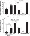
Figure 1
Figure 1
Changes in the number of (a) blood vessels and (b) pericytes in the corpus luteum throughout the menstrual cycle and in early pregnancy. Immunohistochemistry for CD34, a marker of vascular endothelial cells, and for α‐smooth muscle actin (α‐SMA), a marker of pericytes, was carried out on tissue samples obtainef pregnancy). In the tissue sections for the immunohistochemical study of CD34 and α‐SMA, the number of blood vessels or pericytes was determined by counting the number of CD34‐positive vessels or α‐SMA‐positive vessels per unit area in the histological section at 200×, and the number of luteal cells was also counted in the same area. The number of blood vessels or pericytes was expressed per 100 luteal cells. The number of luteal cells in the corpus luteum of the next follicular phase could not be quantified because of cell death of the luteal cells and was expressed as NC (not countable). Tissue samples were obtained from three different patients for each group. Values are mean + standard error.
Figure 2
Figure 2
Double immunostaining for CD34, a marker for vascular endothelial cells (blue), and Ki‐67, a marker of proliferating cells (brown). In the corpus luteum of the mid‐luteal phase (a) there were almost no Ki‐67 positive cells. However, the corpus luteum of pregnancy (b) had some double‐stained cells. These findings suggest that there are some proliferating endothelial cells in the corpus luteum of early pregnancy, but not in the mid‐luteal phase corpus luteum. Bar represents 30 µm.
Figure 3
Figure 3
Double immunostaining for CD34, which is a marker for vascular endothelial cells (blue), and α‐smooth muscle actin, which is a marker of pericytes (brown). Some endothelial cells are surrounded by pericytes (arrows, a), which suggests mature stabilized blood vessels. Double‐stained blood vessels were observed in the corpus luteum of the mid‐luteal phase (c) and in early pregnancy (d), but not in the corpus luteum of the early luteal phase (b). Bar represents 30 µm.
Figure 4
Figure 4
Possible molecular mechanism of angiogenesis in the corpus luteum (CL) during the menstrual cycle and in early pregnancy. In the early luteal phase, vascular endothelial growth factor (VEGF) stimulates angiogenesis under the environment of high angiopoietin‐2 (Ang‐2) expression, but newly formed blood vessels are immature. In the mid‐luteal phase, pericytes are recruited and high angiopoietin‐1 (Ang‐1) stabilizes blood vessels, but angiogenesis is stopped because VEGF expression remains unchanged. During the regression phase, relatively high angiopoietin‐2 expression resulting from a decrease in angiopoietin‐1 destabilizes blood vessels and loss of VEGF action induces blood vessel regression. In the corpus luteum of pregnancy, angiogenesis is activated again because of strong VEGF expression by human chorionic gonadotropin and high angiopoietin‐1 stabilizes blood vessels, suggesting that angiogenesis is accompanied by blood vessel stabilization.

References

    1. Tamura H, Greenwald GS. Angiogenesis and its hormonal control in the corpus luteum of the pregnant rat. Biol Reprod 1987; 36: 1149–1154. - PubMed
    1. Ferrara N, Chen H, Davis‐Smyth T et al Vascular endothelial growth factor is essential for corpus luteum angiogenesis. Nat Med 1998; 4: 336–340. - PubMed
    1. Fraser HM, Dickson SE, Lunn SF et al Suppression of luteal angiogenesis in the primate after neutralization of vascular endothelial growth factor. Endocrinology 2000; 141: 995–1000. - PubMed
    1. Sugino N, Kashida S, Takiguchi S, Karube A, Kato H. Expression of vascular endothelial growth factor and its receptors in the human corpus luteum during the menstrual cycle and in early pregnancy. J Clin Endocrinol Metab 2000; 85: 3919–3924. - PubMed
    1. Kashida S, Sugino N, Takiguchi S et al Regulation and role of vascular endothelial growth factor in the corpus luteum during mid‐pregnancy in rats. Biol Reprod 2001; 64: 317–323. - PubMed