. 2018 Apr 26;16(1):40.
doi: 10.1186/s12958-018-0357-7.

Docosahexaenoic acid (DHA) effects on proliferation and steroidogenesis of bovine granulosa cells

Affiliations

Docosahexaenoic acid (DHA) effects on proliferation and steroidogenesis of bovine granulosa cells

Virginie Maillard et al. Reprod Biol Endocrinol. .

Abstract

Background: Docosahexaenoic acid (DHA) is a n-3 polyunsaturated fatty acid (PUFA) belonging to a family of biologically active fatty acids (FA), which are known to have numerous health benefits. N-3 PUFAs affect reproduction in cattle, and notably directly affect follicular cells. In terms of reproduction in cattle, n-3 PUFA-enriched diets lead to increased follicle size or numbers.

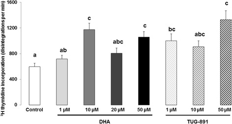
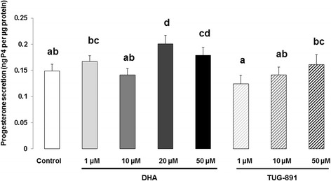
Methods: The objective of the present study was to analyze the effects of DHA (1, 10, 20 and 50 μM) on proliferation and steroidogenesis (parametric and/or non parametric (permutational) ANOVA) of bovine granulosa cells in vitro and mechanisms of action through protein expression (Kruskal-Wallis) and signaling pathways (non parametric ANOVA) and to investigate whether DHA could exert part of its action through the free fatty acid receptor 4 (FFAR4).

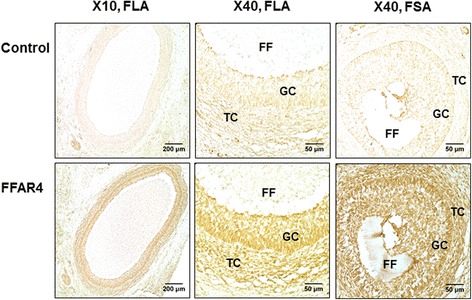
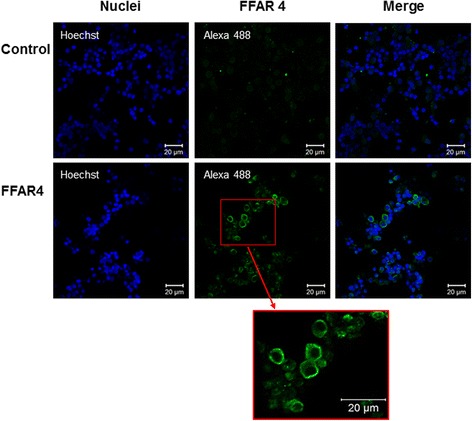
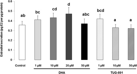
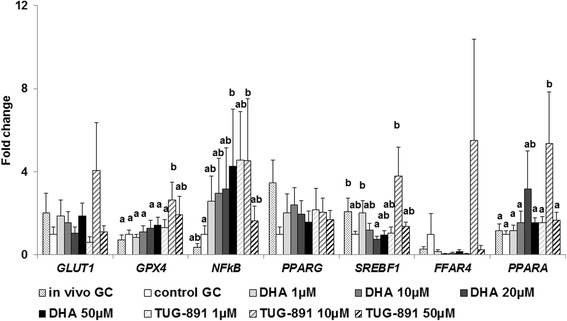
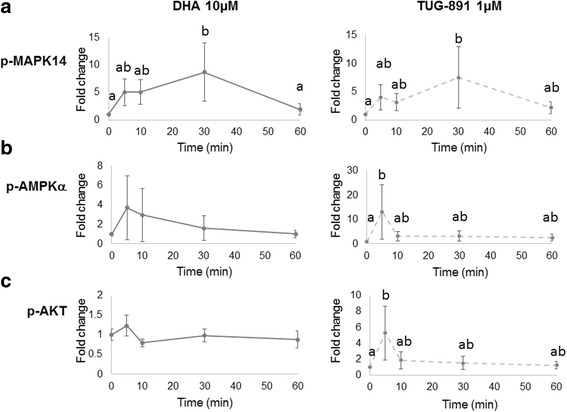
Results: DHA (10 and 50 μM) increased granulosa cell proliferation and DHA 10 μM led to a corresponding increase in proliferating cell nuclear antigen (PCNA) expression level. DHA also increased progesterone secretion at 1, 20 and 50 μM, and estradiol secretion at 1, 10 and 20 μM. Consistent increases in protein levels were also reported for the steroidogenic enzymes, cytochrome P450 family 11 subfamily A member 1 (CYP11A1) and hydroxy-delta-5-steroid dehydrogenase, 3 beta- and steroid delta-isomerase 1 (HSD3B1), and of the cholesterol transporter steroidogenic acute regulatory protein (StAR), which are necessary for production of progesterone or androstenedione. FFAR4 was expressed in all cellular types of bovine ovarian follicles, and in granulosa cells it was localized close to the cellular membrane. TUG-891 treatment (1 and 50 μM), a FFAR4 agonist, increased granulosa cell proliferation and MAPK14 phosphorylation in a similar way to that observed with DHA treatment. However, TUG-891 treatment (1, 10 and 50 μM) showed no effect on progesterone or estradiol secretion.

Conclusions: These data show that DHA stimulated proliferation and steroidogenesis of bovine granulosa cells and led to MAPK14 phosphorylation. FFAR4 involvement in DHA effects requires further investigation, even if our data might suggest FFAR4 role in DHA effects on granulosa cell proliferation. Other mechanisms of DHA action should be investigated as the steroidogenic effects seemed to be independent of FFAR4 activation.

Keywords: AKT; AMPK; Bovine; DHA; Folliculogenesis; Free fatty acid receptor 4 (FFAR4); Gene expression; Lipid; MAPK; N-3 PUFA; Signaling pathways.


Conflict of interest statement

Ethics approval and consent to participate

Not applicable.

Competing interests

The authors declare that they have no competing interests.

Publisher’s Note

Springer Nature remains neutral with regard to jurisdictional claims in published maps and institutional affiliations.

Figures

Fig. 1
Fig. 1
Expression of free fatty acid receptor 4 (FFAR4) in bovine ovarian follicles by immunohistochemistry. Immunohistochemistry was performed on sections of ovarian follicles. FFAR4 (brown labeling) was immunodetected in ovarian follicles with large (FLA) or small (FSA) antrum (customized FFAR4 rabbit antibody, Agro-Bio). Pre-immunized rabbit serum was used as the control with the same secondary antibody as for FFAR4 detection. Bars = 200 μm and 50 μm for 10× and 40× microscope objectives, respectively. FF - follicular fluid, GC – granulosa cells, TC – theca cells
Fig. 2
Fig. 2
Expression and localization of free fatty acid receptor 4 (FFAR4) in bovine granulosa cells by immunofluorescence. Immunofluorescence was performed on granulosa cells (GCs) after in vitro culture. Briefly, recovered GCs after follicle puncture and GC washing were incubated in serum-free modified McCoy’s 5A medium (2.5 × 105 viable cells/well on a 8-well chamber slide (Lab-Tek® Nunc) for 48 h. Cultures were performed in a water-saturated atmosphere containing 5% CO2 in air at 38 °C. FFAR4 (green fluorescence) was immunodetected in GCs (customized FFAR4 rabbit antibody, Agro-Bio). Pre-immunized rabbit serum was used as the control with the same secondary antibody as for FFAR4 detection. Nuclei were stained with Hoechst 33,258 (blue fluorescence). The picture framed in red is a magnification of the area framed in red from the original image. Bars = 20 μm
Fig. 3
Fig. 3
Synthesis of DNA in bovine granulosa cells: 3H-thymidine incorporation after 24 h treatment with DHA or TUG-891. Effects of DHA or TUG-891 on cell proliferation were assessed by measurement of 3H-thymidine incorporation in bovine granulosa cells after 24 h culture in enriched McCoy’s 5A media with various doses of DHA (1, 10, 20 and 50 μM) or TUG-891 (1, 10 and 50 μM), as described in Material and Method section. The chemical DMSO alone (1/2000) was used as a negative control due to its use as a solvent for DHA and TUG-891. The data are expressed as disintegrations per minute. Results represent 13 independent cultures with each treatment conducted in four replicates and are presented as mean ± SEM. Bars with different superscripts are significantly different (p < 0.05)
Fig. 4
Fig. 4
Progesterone secretion from bovine granulosa cells after 48 h treatment with DHA or TUG-891. Effects of DHA or TUG-891 on progesterone secretion were assessed in culture media of bovine granulosa cells cultured for 48 h in enriched McCoy’s 5A media with various doses of DHA (1, 10, 20 and 50 μM) or TUG-891 (1, 10 and 50 μM), as described in Material and Method section. The chemical DMSO alone (1/2000) was used as a negative control due to its use as a solvent for DHA and TUG-891. Progesterone secretion was normalized with the protein concentration in each well and expressed as ng progesterone (P4) per μg protein. Results of 12 independent cultures, with each treatment conducted in quadruplicate, are presented as mean ± SEM. Bars with different superscripts are significantly different (p < 0.05)
Fig. 5
Fig. 5
Estradiol secretion from bovine granulosa cells after 48 h treatment with DHA or TUG-891. Effects of DHA or TUG-891 on estradiol secretion were assessed in culture media of bovine granulosa cells cultured for 48 h in enriched McCoy’s 5A media with various doses of DHA (1, 10, 20 and 50 μM) or TUG-891 (1, 10 and 50 μM), as described in Material and Method section. The chemical DMSO alone (1/2000) was used as a negative control due to its use as a solvent for DHA and TUG-891. Estradiol secretion was normalized with the protein concentration in each well and expressed as pg estradiol (E2) per μg protein. Results of six independent cultures, with each treatment conducted in quadruplicate, are presented as mean ± SEM. Bars with different superscripts are significantly different (p < 0.05)
Fig. 6
Fig. 6
Protein expression of (a) proliferating cell nuclear antigen (PCNA), (b) hydroxy-delta-5-steroid dehydrogenase, 3 beta- and steroid delta-isomerase 1 (HSD3B1), (c) steroidogenic acute regulatory protein (StAR) and (d) cytochrome P450 family 11 subfamily A member 1 (CYP11A1) after 15 h treatment with DHA. Effects of DHA treatment on protein levels were assessed in bovine granulosa cells after 15 h culture in enriched McCoy’s 5A media in presence or absence of DHA 10 or 20 μM. The chemical DMSO alone (1/2000) was used as a negative control due to its solvent activity on DHA. Protein extracts were separated by electrophoresis on 4–12% (w:v) SDS-polyacrylamide gel. After electrotransfer to nitrocellulose membranes, the proteins were probed with anti-PCNA (a), anti-HSD3B1 (b), anti-StAR (c) or anti-CYP11A1 (d) antibodies. The blots were stripped and re-probed with antibodies against Vinculin (VCL). Results of at least five independent experiments are presented. Bands on the blots were quantified and the total protein / VCL protein ratio was calculated. Results are expressed relative to the control as mean ± SEM of five independent experiments for HSD3B1, StAR, CYP11A1 and six independent experiments for PCNA. * indicates significant difference (p < 0.05) and # indicates tendency (p < 0.10)
Fig. 7
Fig. 7
Gene expression in bovine granulosa cells before or after 8 h treatment with DHA or TUG-891. Effects of DHA or TUG-891 on mRNA expression of solute carrier family 2 member 1 (GLUT1), glutathione peroxidase 4 (GPX4), nuclear factor of kappa light polypeptide gene enhancer in B-cells 1 (NFkB), peroxisome proliferator-activated receptor gamma (PPARG), sterol regulatory element binding transcription factor 1 (SREBF1), free fatty acid receptor 4 (FFAR4) and PPAR alpha (PPARA) were assessed in bovine granulosa cells (GC) before or after 8 h culture in enriched McCoy’s 5A media with various doses of DHA (1, 10, 20 and 50 μM) or TUG-891 (1, 10 and 50 μM), as described in Material and Method section. The chemical DMSO alone (1/2000) was used as a negative control due to its use as a solvent for DHA and TUG-891. Total mRNA was extracted from GC and reverse-transcribed, and real-time RT-PCR was performed. The geometric mean of two housekeeping genes (RPL19- ribosomal protein L19 and RPS9- ribosomal protein S9) was used to normalize gene expression. Results of 10 independent cultures are presented as mean ± SEM. Bars with different superscripts are significantly different (p < 0.05)
Fig. 8
Fig. 8
Signaling pathways in bovine granulosa cells after DHA or TUG-891 treatment. Effects of DHA or TUG-891 on phosphorylation of (a) mitogen-activated protein kinase 14 (MAPK14), (b) AMP-activated protein kinaseα (AMPKα) and (c) protein kinase B (Akt) signaling pathways were assessed in bovine granulosa cells cultured for 15 h in enriched McCoy’s 5A media with 10 μM DHA or 1 μM TUG-891, as described in Material and Method section for 5, 10, 30 and 60 min. Protein extracts were separated by electrophoresis on 4–12% (w:v) SDS-polyacrylamide gel. After electrotransfer to nitrocellulose membranes, the proteins were probed with anti-phosphorylated (p-)MAPK14 (a), anti-p-AMPKα (b) or anti-p-AKT1/2/3 (c) antibodies. The blots were stripped and re-probed with antibodies against MAPK14, AMPKα or Akt, respectively. Bands on the blots were quantified. Results of four independent experiments are presented as the ratio of p-protein to total protein, normalized by the ratio observed in control at each time and expressed as mean ± SEM of four independent experiments, with time 0 min being equal to 1 (for reference). Bars with different superscripts are significantly different (p < 0.05)

References

    1. Siriwardhana N, Kalupahana NS, Moustaid-Moussa N, Se-Kwon K. Adv Food Nutr Res. Vol. volume 65: academic press. 2012. Chapter 13 - health benefits of n-3 polyunsaturated fatty acids: Eicosapentaenoic acid and docosahexaenoic acid; pp. 211–222. - PubMed
    1. Plourde M, Cunnane SC. Extremely limited synthesis of long chain polyunsaturates in adults: implications for their dietary essentiality and use as supplements. Appl Physiol Nutr Metab. 2007;32(4):619–634. doi: 10.1139/H07-034. - DOI - PubMed
    1. Baker EJ, Miles EA, Burdge GC, Yaqoob P, Calder PC. Metabolism and functional effects of plant-derived omega-3 fatty acids in humans. Prog Lipid Res. 2016;64:30–56. doi: 10.1016/j.plipres.2016.07.002. - DOI - PubMed
    1. Simopoulos AP. Omega-3 fatty acids in inflammation and autoimmune diseases. J Am Coll Nutr. 2002;21(6):495–505. doi: 10.1080/07315724.2002.10719248. - DOI - PubMed
    1. Deckelbaum RJ, Torrejon C. The omega-3 fatty acid nutritional landscape: health benefits and sources. J Nutr. 2012;142(3):587S–591S. doi: 10.3945/jn.111.148080. - DOI - PMC - PubMed

Substances

LinkOut - more resources