

Cardiac amyloidosis
- PMID: 29700090
- PMCID: PMC6334035
- DOI: 10.7861/clinmedicine.18-2-s30
Cardiac amyloidosis
Abstract
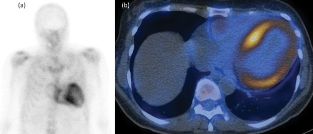
Systemic amyloidosis comprises an uncommon group of disorders caused by the extracellular deposition of misfolded proteins in various organs. Cardiac amyloid deposition, causing an infiltrative/restrictive cardiomyopathy, is a frequent feature of amyloidosis and a major determinant of survival. It may be the presenting feature of the disease or may be identified while investigating a patient presenting with other organ involvement. The need for a high index of suspicion and the critical importance of precise biochemical typing of the amyloid deposits is paramount in light of recent therapeutic advances that can significantly improve prognosis. Most cases of cardiac amyloidosis are of either transthyretin type, which may be acquired in older individuals or inherited in younger patients, or acquired monoclonal immunoglobulin light chain (AL) type. This article aims to review recent developments in diagnosis and management of cardiac amyloidosis.
Keywords: AL amyloidosis; ATTR amyloidosis; CMR; cardiac amyloidosis; infiltrative cardiomyopathy.
© Royal College of Physicians 2018. All rights reserved.
Figures
References
-
- Lachmann HJ. Hawkins PN. Systemic amyloidosis. Curr Opin Pharmacol. 2006;6:214–20. - PubMed
-
- Sipe JD. Benson MD. Buxbaum JN, et al. Nomenclature 2014: Amyloid fibril proteins and clinical classification of the amyloidosis. Amyloid. 2014;21:221–4. - PubMed
-
- Fontana M. Banypersad SM. Treibel TA, et al. Differential myocyte responses in patients with cardiac transthyretin amyloidosis and light-chain amyloidosis: a cardiac MR imaging study. Radiology. 2015;277:388–97. - PubMed
-
- Falk RH. Diagnosis and management of the cardiac amyloidoses. Circulation. 2005;112:2047–60. - PubMed
-
- Rapezzi C. Merlini G. Quarta CC, et al. Systemic cardiac amyloidoses: disease profiles and clinical courses of the 3 main types. Circulation. 2009;120:1203–12. - PubMed
Publication types
MeSH terms
LinkOut - more resources
Full Text Sources
Other Literature Sources
Medical
Research Materials
