. 2018 Jun 8;293(23):8969-8981.
doi: 10.1074/jbc.RA117.001167. Epub 2018 Apr 26.

COX-2 expression mediated by calcium-TonEBP signaling axis under hyperosmotic conditions serves osmoprotective function in nucleus pulposus cells

Affiliations

COX-2 expression mediated by calcium-TonEBP signaling axis under hyperosmotic conditions serves osmoprotective function in nucleus pulposus cells

Hyowon Choi et al. J Biol Chem. .

Abstract

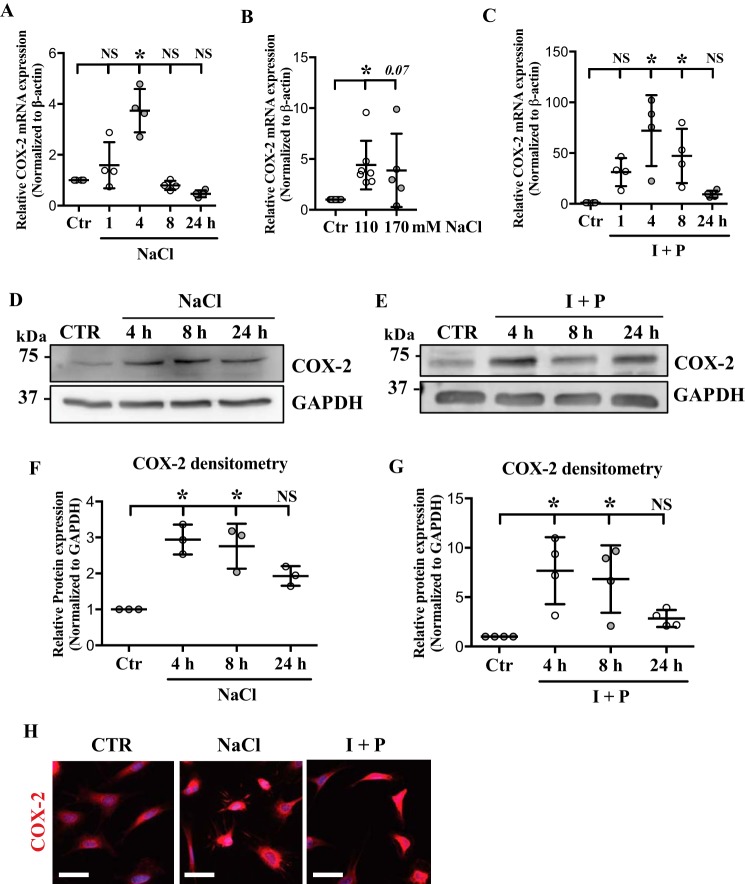
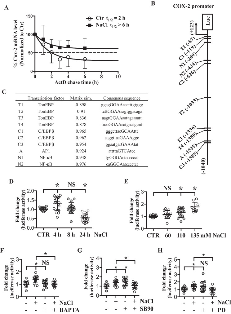
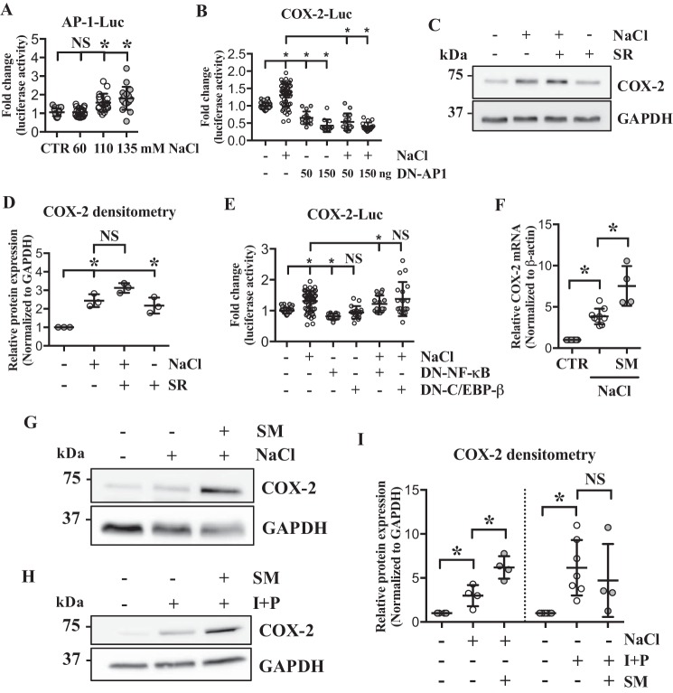
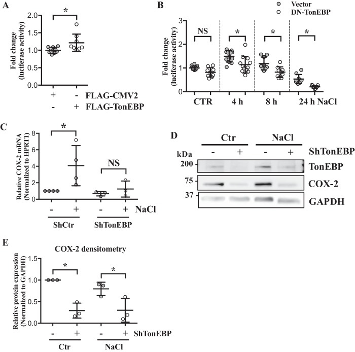
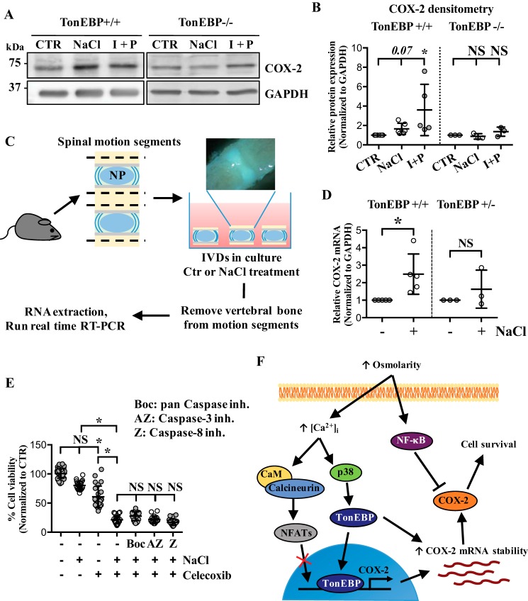
The nucleus pulposus (NP) of intervertebral discs experiences dynamic changes in tissue osmolarity because of diurnal loading of the spine. TonEBP/NFAT5 is a transcription factor that is critical in osmoregulation as well as survival of NP cells in the hyperosmotic milieu. The goal of this study was to investigate whether cyclooxygenase-2 (COX-2) expression is osmoresponsive and dependent on TonEBP, and whether it serves an osmoprotective role. NP cells up-regulated COX-2 expression in hyperosmotic media. The induction of COX-2 depended on elevation of intracellular calcium levels and p38 MAPK pathway, but independent of calcineurin signaling as well as MEK/ERK and JNK pathways. Under hyperosmotic conditions, both COX-2 mRNA stability and its proximal promoter activity were increased. The proximal COX-2 promoter (-1840/+123 bp) contained predicted binding sites for TonEBP, AP-1, NF-κB, and C/EBP-β. While COX-2 promoter activity was positively regulated by both AP-1 and NF-κB, AP-1 had no effect and NF-κB negatively regulated COX-2 protein levels under hyperosmotic conditions. On the other hand, TonEBP was necessary for both COX-2 promoter activity and protein up-regulation in response to hyperosmotic stimuli. Ex vivo disc organ culture studies using hypomorphic TonEBP+/- mice confirmed that TonEBP is required for hyperosmotic induction of COX-2. Importantly, the inhibition of COX-2 activity under hyperosmotic conditions resulted in decreased cell viability, suggesting that COX-2 plays a cytoprotective and homeostatic role in NP cells for their adaptation to dynamically loaded hyperosmotic niches.

Keywords: COX-2; NFAT transcription factor; TonEBP; calcium; cell biology; cell signaling; cyclooxygenase (COX); intervertebral disc; nucleus pulposus; osmoregulation; transcription factor.


Conflict of interest statement

The authors declare that they have no conflicts of interest with the contents of this article

Figures

Figure 1.
Figure 1.
COX-2 is up-regulated in response to hyperosmolarity and high intracellular calcium in NP cells. A and B, qRT-PCR analysis demonstrates (A) time- and (B) dose-dependent COX-2 mRNA increase under hyperosmotic conditions. C, COX-2 mRNA is induced by ionomycin/PMA (I + P) treatment. D and E, Western blots of COX-2 after NaCl or I + P time course treatments show significant induction of COX-2 expression. F and G, densitometry analyses of Western blots from D and E. H, immunofluorescence staining of COX-2 in NP cells confirms that its expression is increased with NaCl as well as I + P treatment. Scale bar: 50 μm. All the quantitative data are represented as mean ± S.D. from at least three independent experiments. *, p < 0.05.
Figure 2.
Figure 2.
Hyperosmolarity-mediated induction of COX-2 is through intracellular calcium but independent of calcineurin signaling. A–C, hyperosmotic induction of COX-2 mRNA (A), and protein (B and C) is suppressed by BAPTA, a calcium chelator, but is unaffected by FK-506/CsA (F/C), a calcineurin inhibitor. D–F, induction of COX-2 mRNA (D) and protein (E and F) in response to ionomycin/PMA (I + P) treatment is completely abolished with BAPTA, but not with FK/CsA treatment. All the quantitative data are represented as mean ± S.D. from at least three independent experiments (three biological replicates). NS: nonsignificant; *, p < 0.05.
Figure 3.
Figure 3.
Hyperosmotic induction of COX-2 is mediated by p38, but not by ERK or JNK. A and B, phospho-p38 (p-p38) levels significantly increase in response to hyperosmolarity. C and D, hyperosmotic increase of COX-2 mRNA is completely suppressed by p38 inhibitor, SB202190, but unaffected by JNK inhibitor, SP600125, and MEK/ERK inhibitor, PD98059. E, Western blot images showing that p38 inhibition prevents COX-2 induction in response to hyperosmolarity. F, Western blot images showing that ionomycin/PMA (I + P)-mediated COX-2 induction is not affected by p38 inhibition. G, densitometry analyses of Western blots shown in E and F. All the quantitative data are represented as mean ± S.D. from at least three independent experiments (three biological replicates). NS: nonsignificant; *, p < 0.05. PD: PD98059 (MEK/ERK inhibitor); SB90: SB202190 (p38 inhibitor); SP: SP600125 (JNK inhibitor).
Figure 4.
Figure 4.
Both increased mRNA stability and proximal promoter activity account for COX-2 up-regulation in response to hyperosmolarity. A, actinomycin D chase study demonstrates that estimated half-life of COX-2 mRNA is increased by hyperosmolarity, representing increased mRNA stability (n = 5). B, a schematic showing TonEBP, AP-1, NF-κB, and C/EBP-β–binding sites on ∼1.8 kb COX-2 proximal promoter region. C, list of putative binding sites and consensus sequence for each transcription factor. Four bp core consensus sequence used for query was marked as capital letters. Matrix similarity score based on the most conserved nucleotide at each position of the matrix is also shown. D and E, COX-2 promoter activity shows (D) time- and (E) dose-dependent increase under hyperosmotic conditions. F, hyperosmolarity-dependent increase of COX-2 promoter activity is inhibited with a calcium chelator, BAPTA. G and H, up-regulation of COX-2 promoter activity under hyperosmotic condition was inhibited by p38 inhibitor (SB90) and MEK/ERK inhibitor (PD). All the quantitative data are represented as mean ± S.D. from at least three independent experiments (three biological replicates). Promoter activity experiments were done with three technical replicates per independent experiment. NS: nonsignificant; *, p < 0.05. SB90: SB202190 (p38 inhibitor); PD: PD98059 (MEK/ERK inhibitor).
Figure 5.
Figure 5.
AP-1, NF-κB, and C/EBP-β are not involved in hyperosmotic induction of COX-2. A, luciferase assay using AP-1 reporter shows that AP-1 activity is up-regulated by hyperosmolarity. B, COX-2 promoter activity under hyperosmotic condition is significantly decreased by DN–AP-1. C and D, AP-1 inhibition by a specific inhibitor, SR11302, does not block hyperosmotic induction of COX-2. E, COX-2 promoter activity is decreased by DN–NF-κB, but unaltered by DN–C/EBP-β. F, unlike the promoter activities, COX-2 mRNA levels are further up-regulated by NF-κB inhibitor, SM7368, under hyperosmotic condition. G–I, NF-κB inhibition resulted in further up-regulation of COX-2 protein levels under hyperosmotic condition, but had no effect when the cells were treated with ionomycin/PMA. All the quantitative data are represented as mean ± S.D. from at least three independent experiments (three biological replicates). Promoter activity experiments were done with three technical replicates per independent experiment. NS: nonsignificant; *, p < 0.05. SR: SR11302 (AP-1 inhibitor); SM: SM7368 (NF-κB inhibitor); I + P: ionomycin/PMA.
Figure 6.
Figure 6.
TonEBP is necessary for hyperosmotic induction of COX-2 in NP cells. A, COX-2 promoter activity is significantly increased by TonEBP overexpression. B, transfection of cells with DN–TonEBP under hyperosmotic conditions resulted in significant decrease in COX-2 promoter activity. C, stable silencing of TonEBP results in inhibition of COX-2 mRNA induction in response to hyperosmolarity. D and E, Western blotting and densitometry analyses of COX-2 show that without TonEBP, NP cells are unable to up-regulate COX-2 under hyperosmotic condition. All the quantitative data are represented as mean ± S.D. from at least three independent experiments (three biological replicates). Promoter activity experiments were done with three technical replicates per independent experiment. NS: nonsignificant; *, p < 0.05.
Figure 7.
Figure 7.
COX-2 activity under hyperosmotic condition promotes NP cell survival. A and B, TonEBP null MEFs, unlike WT MEFs, do not induce COX-2 in response to either hyperosmolarity or ionomycin/PMA treatment. C, a schematic describing ex vivo disc organ culture system. Briefly, mouse disc motion segments were dissected from WT or haploinsufficient TonEBP+/− mice and cultured in isoosmotic or hyperosmotic media, and then tissue RNA was extracted to perform qRT-PCR. Picture in the schematic shows single motion segment. D, TonEBP+/− mouse discs do not up-regulate COX-2 mRNA in response to hyperosmotic stimulus. E, MTT assay demonstrates a significant reduction in cell viability with celecoxib, a COX-2 inhibitor, under both isoosmotic and hyperosmotic conditions. This cell death cannot be rescued by various caspase inhibitors. F, increased extracellular osmolarity leads to increased intracellular calcium levels, which in turns controls various cellular pathways including p38 MAPK to activate TonEBP. Although intracellular calcium can activate calcineurin signaling pathway, calcineurin-NFAT pathway is not involved in hyperosmotic up-regulation of COX-2 in NP cells. TonEBP transcriptionally up-regulates COX-2 expression and increases COX-2 mRNA stability, eventually promoting NP cell survival under hyperosmotic condition. In contrast, NF-κB negatively regulates COX-2 protein expression under hyperosmotic condition. All the quantitative data are represented as mean ± S.D. from at least three independent experiments (three biological replicates). Cell viability experiments were performed with four technical replicates per independent experiment. NS: nonsignificant; *, p < 0.05. I + P: ionomycin/PMA; Boc: Boc-D-FMK (pan caspase inhibitor), AZ: AZ10417808 (caspase-3 inhibitor); Z: Z-IE(OMe)TD(OMe)-FMK (caspase-8 inhibitor).

References

    1. Johnson Z. I., Shapiro I. M., and Risbud M. (2014) Extracellular osmolarity regulates matrix homeostasis in the intervertebral disc and articular cartilage: Evolving role of TonEBP. Matrix Biol. 40, 10–16 10.1016/j.matbio.2014.08.014 - DOI - PMC - PubMed
    1. Silagi E. S., Shapiro I. M., and Risbud M. V. (2018) Glycosaminoglycan synthesis in the nucleus pulposus: Dysregulation and the pathogenesis of disc degeneration. Matrix Biol. 10.1016/j.matbio.2018.02.025 - DOI - PMC - PubMed
    1. Binch A. L. A., Shapiro I. M., and Risbud M. V. (2016) Syndecan-4 in intervertebral disc and cartilage: Saint or synner? Matrix Biol. 52-54, 355–362 10.1016/j.matbio.2016.01.005 - DOI - PMC - PubMed
    1. Risbud M. V., and Shapiro I. M. (2014) Role of cytokines in intervertebral disc degeneration: Pain and disc content. Nat. Rev. Rheumatol. 10, 44–56 10.1038/nrrheum.2013.160 - DOI - PMC - PubMed
    1. Choi H., Tessier S., Silagi E. S., Kyada R., Yousefi F., Pleshko N., Shapiro I. M., and Risbud M. V. (2018) A novel mouse model of intervertebral disc degeneration shows altered cell fate and matrix homeostasis. Matrix Biol. 10.1016/j.matbio.2018.03.019 - DOI - PMC - PubMed

Publication types

MeSH terms