. 2018 Apr 27:23:274-284.
doi: 10.12659/AOT.907796.

Anesthesia Management of Modified Ex Vivo Liver Resection and Autotransplantation

Affiliations

Anesthesia Management of Modified Ex Vivo Liver Resection and Autotransplantation

Fujun Cheng et al. Ann Transplant. .

Abstract

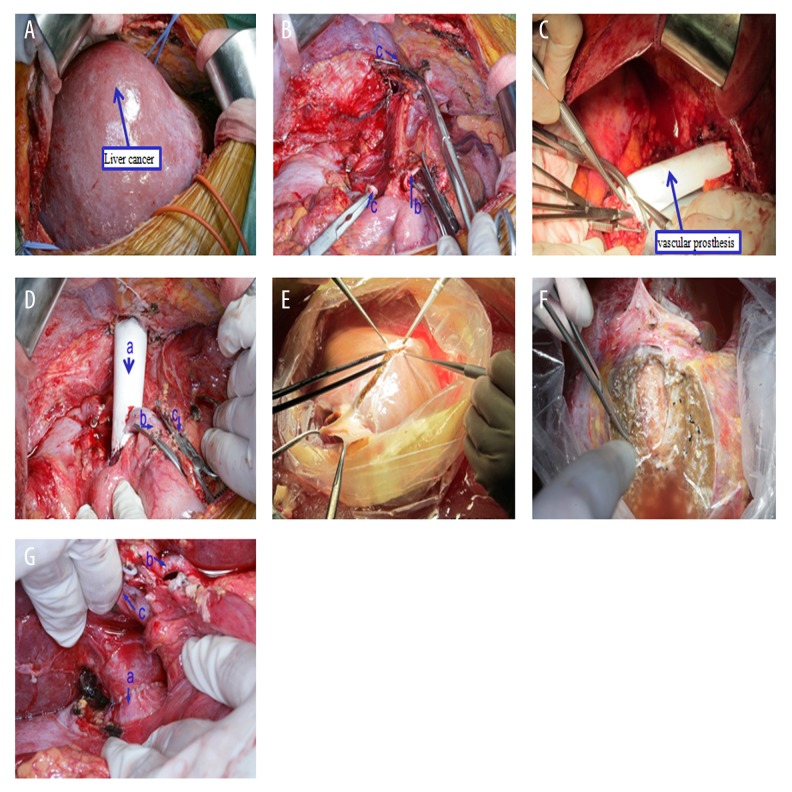
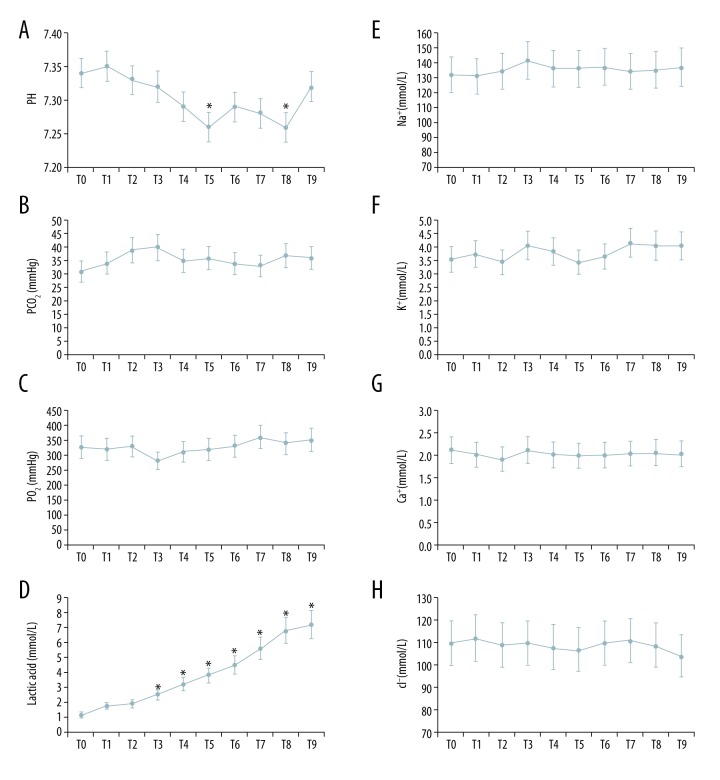
BACKGROUND Ex situ liver surgery allows liver resection and vascular reconstruction in patients who have liver tumors located in critical sites. Only a small series of studies about ex situ liver surgery is available in the literature. No anesthesia management experience has been previously published. The aim of the currents study was to summarize our experience with anesthetic management of patients during ex vivo liver surgery. MATERIAL AND METHODS The first 43 patients who received ex vivo liver surgery between January 2007 and April 2012 were included. A pulmonary artery catheter (PAC), transesophageal echocardiography (TEE), and pulse indicator continuous cardiac output (PiCCO) were used intraoperatively in the patients to monitor the hemodynamic changes. Thromboelastogram and the plasma coagulation test were used to monitor the coagulation changes. RESULTS All patients received general anesthesia with rapid sequence induction. The data obtained by PAC, TEE, and PiCOO in these cases showed large changes in hemodynamics during the stages of the first or second vessel reconstruction. The CI decreased about 59%/63% and the MPAP decreased about 49%/37% during the first/second vessel reconstruction. Accurate judgment of the dosage of active drug for vascular support is the key for the stabilization of hemodynamics as quickly as possible. However, a high incidence (35.5%) of prophase fibrinolysis in a long anhepatic phase should be monitored and managed. CONCLUSIONS Ex vivo liver surgery is no longer experimental and is a therapeutic option for patients with liver cancer in critical sites. Good anesthesia support is an essential element of liver autotransplantation.


Figures

Figure 1
Figure 1
The images show the typical surgery procedures of a modified ex situ ex vivo liver resection and autotransplantation. (A) The large convex lump in the top portion of the liver was liver cancer; (B) The first porta hepatis structures after the liver were removed from the body; (C) The inferior vena cava was rebuilt with a vascular prosthesis; (D) The portal vein was anastomosed with the vascular prosthesis; (E) Ex situ ex vivo liver cancer resection in the HTK preservative fluid; (F) The liver cancer was resected from the liver, and the vessel stumps were ligatured; (G) The porta hepatis structures after the liver were autotransplanted. a: inferior vena cava or vascular prosthesis; b: hepatic artery; c: portal vein.
Figure 2
Figure 2
Hemodynamic data (mean ±SD, n=31) measured by pulmonary artery catheter, TEE, and PiCCO. (A) Heart rate (HR); (B) Mean arterial pressure (MAP); (C) Central vascular pressure (CVP); (D) Cardiac index (CI); (E) Pulmonary vascular resistance index (PVRI); (F) Systemic vascular resistance index (SVRI); (G) Mean pulmonary arterial pressure (MPAP); (H) Stroke volume (SV); (I) Pulmonary capillary wedge pressure (PCWP); (J) Vasoactive agents were used during operation. T0: after intubation and cannulation; T1: 5 min before the first vessel reconstruction; T2: immediately after starting the first vessel reconstruction; T3: 5 min after starting the first vessel reconstruction; T4: 5 min after the first reperfusion; T5: 5 min before the second vessel reconstruction; T6: immediately after starting the second vessel reconstruction; T7: 5 min after starting the second vessel reconstruction; T8, 5 min after the second reperfusion; and T9: at the end of the surgery. P<0.05 vs. baseline (T0).
Figure 3
Figure 3
Coagulation changes measured by thromboelastogram. (A) Coagulation index (CI); (B) Reaction time (R); (C) K time, the sludged blood formative time (K); (D) α angle (α); (E) Maximum amplitude (Ma); (F) Whole-blood clot lysis index at 30 min (CL30); (G) Fibrinolysis cases: T0: after intubation and cannulation; T1: 5 min before the first vessel reconstruction; T3: 5 min after starting the first vessel reconstruction; T5: 5 min before the second vessel reconstruction; T7: 5 min after starting the second vessel reconstruction; and T9: at the end of the surgery. P<0.05 vs. baseline (T0).
Figure 4
Figure 4
Coagulation changes measured by the plasma coagulation test. (A) Thrombin time (TT); (B) Fibrinogen (Fib); (C) Activated coagulation time of whole blood (APTT); (D) Prothrombin time (PT). T0: after intubation and cannulation; T1: 5 min before the first vessel reconstruction; T3: 5 min after starting the first vessel reconstruction; T5: 5 min before the second vessel reconstruction; T7: 5 min after starting the second vessel reconstruction; and T9: at the end of the surgery. P<0.05 vs. baseline (T0).
Figure 5
Figure 5
The changes in electrolytes and blood gas analysis. (A) Hydrogen-Ion concentration (PH); (B) Partial pressure of carbon dioxide (PCO2); (C) Partial pressure of oxygen (PO2); (D) Lactic acid concentration; (E) Natrium ion concentration (Na+); (F) Potassium ion concentration (K+); (G) Calcium ion concentration (Ca2+); (H) Chlorine ion concentration (cl). T0: after intubation and cannulation; T1: 5 min before the first vessel reconstruction; T2: immediately after starting the first vessel reconstruction; T3: 5 min after starting the first vessel reconstruction; T4: 5 min after the first reperfusion; T5: 5 min before the second vessel reconstruction; T6: immediately after starting the second vessel reconstruction; T7: 5 min after starting the second vessel reconstruction; T8: 5 min after the second reperfusion; and T9: at the end of the surgery. P<0.05 vs. baseline (T0).

References

    1. Jawan B, Wang CH, Chen CL, et al. Review of anesthesia in liver transplantation. Acta Anaesthesiol Taiwan. 2014;52(4):185–96. - PubMed
    1. Oldhafer KJ, Lang H, Schlitt HJ, et al. Long-term experience after ex situ liver surgery. Surgery. 2000;127(5):520–27. - PubMed
    1. Chui A. Ex situ ex vivo liver resection, partial liver autotransplantation for advanced hilar cholangiocarcinoma: A case report. Transplant Proc. 2003;35(1):402–3. - PubMed
    1. Vicente E, Quijano Y, Ielpo B, et al. Ex situ hepatectomy and liver autotransplantation for cholangiocarcinoma. Ann Surg Oncol. 2017;24(13):3990. - PubMed
    1. Baker MA, Maley WR, Needleman L, Doria C. Ex vivo resection of hepatic neoplasia and autotransplantation: A case report and review of the literature. J Gastrointest Surg. 2015;19(6):1169–76. - PubMed

Substances