

A unique hybrid-structured surface produced by rapid electrochemical anodization enhances bio-corrosion resistance and bone cell responses of β-type Ti-24Nb-4Zr-8Sn alloy
- PMID: 29700340
- PMCID: PMC5920132
- DOI: 10.1038/s41598-018-24590-x
A unique hybrid-structured surface produced by rapid electrochemical anodization enhances bio-corrosion resistance and bone cell responses of β-type Ti-24Nb-4Zr-8Sn alloy
Abstract
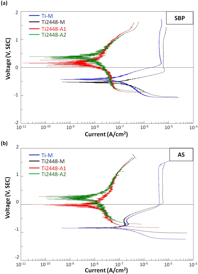
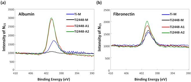
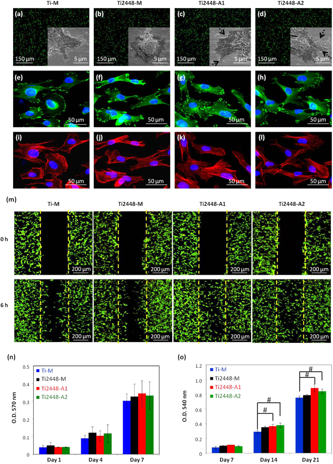
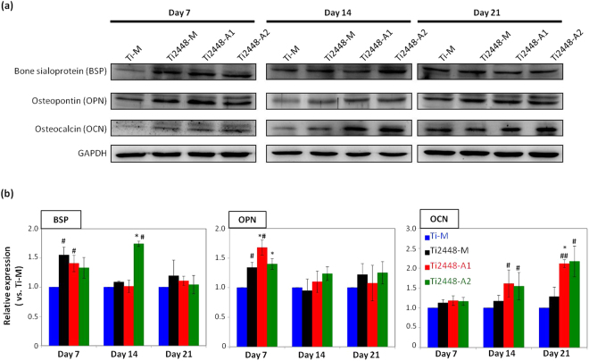
Ti-24Nb-4Zr-8Sn (Ti2448), a new β-type Ti alloy, consists of nontoxic elements and exhibits a low uniaxial tensile elastic modulus of approximately 45 GPa for biomedical implant applications. Nevertheless, the bio-corrosion resistance and biocompatibility of Ti2448 alloys must be improved for long-term clinical use. In this study, a rapid electrochemical anodization treatment was used on Ti2448 alloys to enhance the bio-corrosion resistance and bone cell responses by altering the surface characteristics. The proposed anodization process produces a unique hybrid oxide layer (thickness 50-120 nm) comprising a mesoporous outer section and a dense inner section. Experiment results show that the dense inner section enhances the bio-corrosion resistance. Moreover, the mesoporous surface topography, which is on a similar scale as various biological species, improves the wettability, protein adsorption, focal adhesion complex formation and bone cell differentiation. Outside-in signals can be triggered through the interaction of integrins with the mesoporous topography to form the focal adhesion complex and to further induce osteogenic differentiation pathway. These results demonstrate that the proposed electrochemical anodization process for Ti2448 alloys with a low uniaxial tensile elastic modulus has the potential for biomedical implant applications.
Conflict of interest statement
The authors declare no competing interests.
Figures


Similar articles
-
Modified surface morphology of a novel Ti-24Nb-4Zr-7.9Sn titanium alloy via anodic oxidation for enhanced interfacial biocompatibility and osseointegration.Colloids Surf B Biointerfaces. 2016 Aug 1;144:265-275. doi: 10.1016/j.colsurfb.2016.04.020. Epub 2016 Apr 13. Colloids Surf B Biointerfaces. 2016. PMID: 27100853
-
Corrosion and Biological Behaviors of Biomedical Ti-24Nb-4Zr-8Sn Alloy under an Oxidative Stress Microenvironment.ACS Appl Mater Interfaces. 2024 Apr 17;16(15):18503-18521. doi: 10.1021/acsami.4c00562. Epub 2024 Apr 3. ACS Appl Mater Interfaces. 2024. PMID: 38570902
-
Comparison of the osteoblastic activity of low elastic modulus Ti-24Nb-4Zr-8Sn alloy and pure titanium modified by physical and chemical methods.Mater Sci Eng C Mater Biol Appl. 2020 Aug;113:111018. doi: 10.1016/j.msec.2020.111018. Epub 2020 Apr 25. Mater Sci Eng C Mater Biol Appl. 2020. PMID: 32487417
-
Nanotubular surface modification of metallic implants via electrochemical anodization technique.Int J Nanomedicine. 2014 Sep 17;9:4421-35. doi: 10.2147/IJN.S65866. eCollection 2014. Int J Nanomedicine. 2014. PMID: 25258532 Free PMC article. Review.
-
Frontiers and Challenges in Electrochemical Corrosion Monitoring; Surface and Downhole Applications.Sensors (Basel). 2020 Nov 18;20(22):6583. doi: 10.3390/s20226583. Sensors (Basel). 2020. PMID: 33217977 Free PMC article. Review.
Cited by
-
Surface Engineering Strategies to Enhance the In Situ Performance of Medical Devices Including Atomic Scale Engineering.Int J Mol Sci. 2021 Oct 30;22(21):11788. doi: 10.3390/ijms222111788. Int J Mol Sci. 2021. PMID: 34769219 Free PMC article. Review.
-
Surface Modification of Biomedical Ti and Ti Alloys: A Review on Current Advances.Materials (Basel). 2022 Feb 25;15(5):1749. doi: 10.3390/ma15051749. Materials (Basel). 2022. PMID: 35268983 Free PMC article. Review.
-
Assessing the Potential Association Between Microbes and Corrosion of Intra-Oral Metallic Alloy-Based Dental Appliances Through a Systematic Review of the Literature.Front Bioeng Biotechnol. 2021 Mar 15;9:631103. doi: 10.3389/fbioe.2021.631103. eCollection 2021. Front Bioeng Biotechnol. 2021. PMID: 33791285 Free PMC article.
-
Corrosion of Metallic Biomaterials: A Review.Materials (Basel). 2019 Jan 28;12(3):407. doi: 10.3390/ma12030407. Materials (Basel). 2019. PMID: 30696087 Free PMC article. Review.
-
Three-Dimensionally Printed Ti2448 With Low Stiffness Enhanced Angiogenesis and Osteogenesis by Regulating Macrophage Polarization via Piezo1/YAP Signaling Axis.Front Cell Dev Biol. 2021 Nov 15;9:750948. doi: 10.3389/fcell.2021.750948. eCollection 2021. Front Cell Dev Biol. 2021. PMID: 34869337 Free PMC article.
References
-
- Niinomi M, et al. Development of low rigidity β-type titanium alloy for biomedical applications. Mater Trans. 2002;43:2970–7. doi: 10.2320/matertrans.43.2970. - DOI
-
- Niinomi M. Mechanical properties of biomedical titanium alloys. Mater Sci Eng A Struct Mater. 1998;243:231–6. doi: 10.1016/S0921-5093(97)00806-X. - DOI
Publication types
MeSH terms
Substances
LinkOut - more resources
Full Text Sources
Other Literature Sources
