

Reconstructing the Neanderthal brain using computational anatomy
- PMID: 29700382
- PMCID: PMC5919901
- DOI: 10.1038/s41598-018-24331-0
Reconstructing the Neanderthal brain using computational anatomy
Abstract
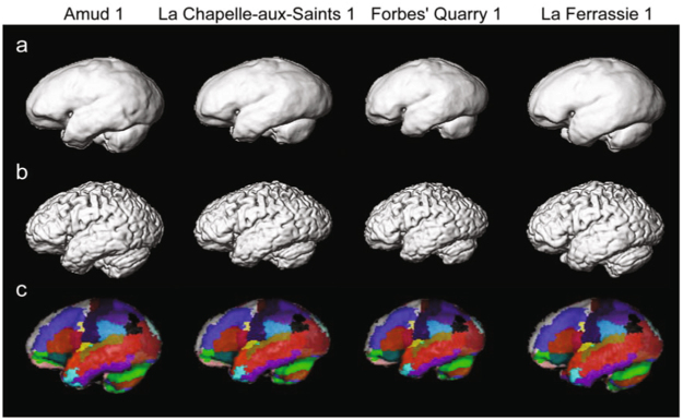
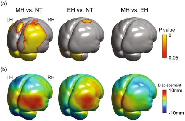
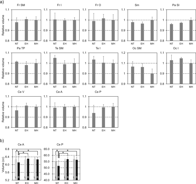
The present study attempted to reconstruct 3D brain shape of Neanderthals and early Homo sapiens based on computational neuroanatomy. We found that early Homo sapiens had relatively larger cerebellar hemispheres but a smaller occipital region in the cerebrum than Neanderthals long before the time that Neanderthals disappeared. Further, using behavioural and structural imaging data of living humans, the abilities such as cognitive flexibility, attention, the language processing, episodic and working memory capacity were positively correlated with size-adjusted cerebellar volume. As the cerebellar hemispheres are structured as a large array of uniform neural modules, a larger cerebellum may possess a larger capacity for cognitive information processing. Such a neuroanatomical difference in the cerebellum may have caused important differences in cognitive and social abilities between the two species and might have contributed to the replacement of Neanderthals by early Homo sapiens.
Conflict of interest statement
The authors declare no competing interests.
Figures
Similar articles
-
Shanidar 3 'rings the bell': Virtual ribcage reconstruction and its implications for understanding the Neanderthal bauplan.J Hum Evol. 2025 Feb;199:103629. doi: 10.1016/j.jhevol.2024.103629. Epub 2024 Dec 11. J Hum Evol. 2025. PMID: 39667186
-
The Neanderthal lower arm.J Hum Evol. 2011 Oct;61(4):396-410. doi: 10.1016/j.jhevol.2011.05.007. Epub 2011 Jul 16. J Hum Evol. 2011. PMID: 21762953
-
Dentine morphology of Atapuerca-Sima de los Huesos lower molars: Evolutionary implications through three-dimensional geometric morphometric analysis.Am J Phys Anthropol. 2018 Jun;166(2):276-295. doi: 10.1002/ajpa.23428. Epub 2018 Feb 8. Am J Phys Anthropol. 2018. PMID: 29417989
-
The false dichotomy: a refutation of the Neandertal indistinguishability claim.J Anthropol Sci. 2016 Jun 20;94:201-21. doi: 10.4436/JASS.94022. Epub 2015 Dec 21. J Anthropol Sci. 2016. PMID: 26708102 Review.
-
Morphology, paleoanthropology, and Neanderthals.Anat Rec. 1998 Aug;253(4):113-7. doi: 10.1002/(SICI)1097-0185(199808)253:4<113::AID-AR6>3.0.CO;2-U. Anat Rec. 1998. PMID: 9740034 Review.
Cited by
-
Keeping 21st Century Paleontology Grounded: Quantitative Genetic Analyses and Ancestral State Reconstruction Re-Emphasize the Essentiality of Fossils.Biology (Basel). 2022 Aug 13;11(8):1218. doi: 10.3390/biology11081218. Biology (Basel). 2022. PMID: 36009845 Free PMC article.
-
Evolutionary and genomic perspectives of brain aging and neurodegenerative diseases.Prog Brain Res. 2023;275:165-215. doi: 10.1016/bs.pbr.2022.10.004. Epub 2023 Feb 3. Prog Brain Res. 2023. PMID: 36841568 Free PMC article.
-
Resistance, vulnerability and resilience: A review of the cognitive cerebellum in aging and neurodegenerative diseases.Neurobiol Learn Mem. 2020 Apr;170:106981. doi: 10.1016/j.nlm.2019.01.004. Epub 2019 Jan 7. Neurobiol Learn Mem. 2020. PMID: 30630042 Free PMC article. Review.
-
Icex: Advances in the automatic extraction and volume calculation of cranial cavities.J Anat. 2023 Jun;242(6):1172-1183. doi: 10.1111/joa.13843. Epub 2023 Feb 11. J Anat. 2023. PMID: 36774197 Free PMC article.
-
Are endocasts good proxies for brain size and shape in archosaurs throughout ontogeny?J Anat. 2019 Mar;234(3):291-305. doi: 10.1111/joa.12918. Epub 2018 Dec 3. J Anat. 2019. PMID: 30506962 Free PMC article.
References
-
- Van Andel, T. H. & Davies, W. Neanderthals and Modern humans in the European Landscape During the Last Glaciation: Archaological Results of the Stage 3 Project. (McDonald Institute for Archaological Research, Cambridge, 2003).
-
- Shea JJ. Transitions or turnovers? Climatically-forced extinctions of Homo sapiens and Neanderthals in the east Mediterranean. Quat. Sci. Rev. 2008;27:2253–2270. doi: 10.1016/j.quascirev.2008.08.015. - DOI
Publication types
MeSH terms
Grants and funding
LinkOut - more resources
Full Text Sources
Other Literature Sources
Molecular Biology Databases
Research Materials
