. 2018 Apr 26;8(1):6580.
doi: 10.1038/s41598-018-24814-0.

Identification of the allosteric P2X7 receptor antagonist [11C]SMW139 as a PET tracer of microglial activation

Affiliations

Identification of the allosteric P2X7 receptor antagonist [11C]SMW139 as a PET tracer of microglial activation

Bieneke Janssen et al. Sci Rep. .

Abstract

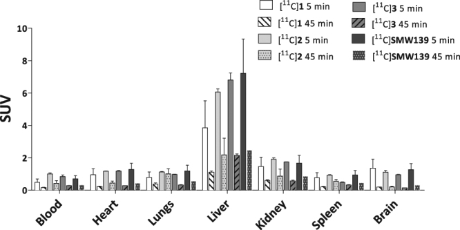
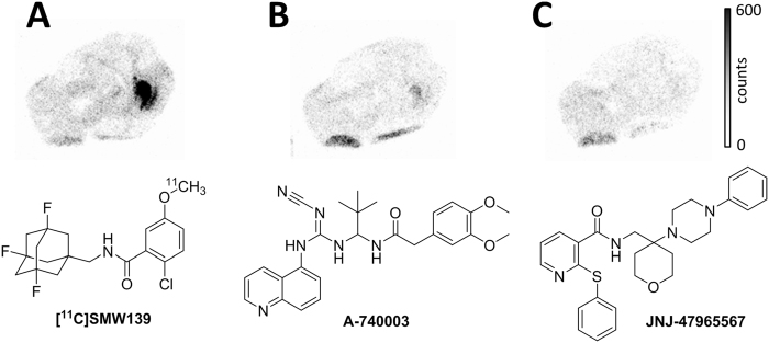
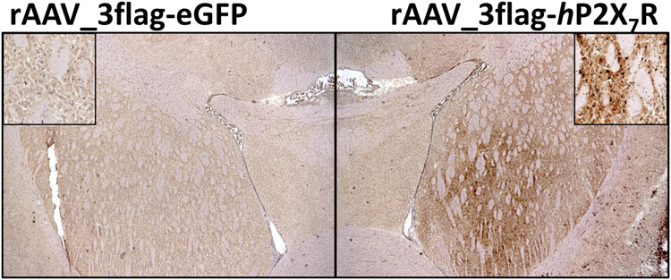
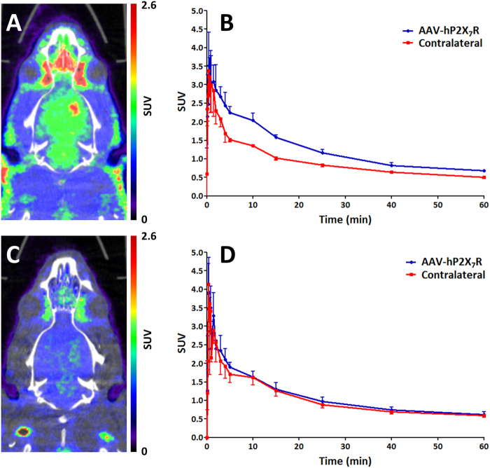
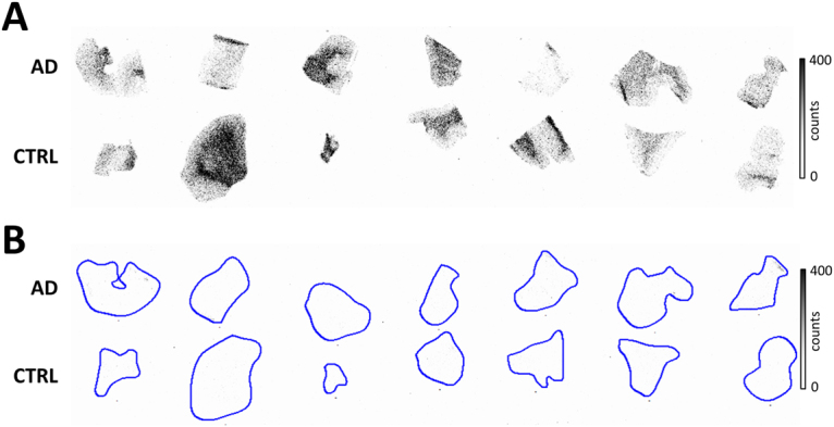
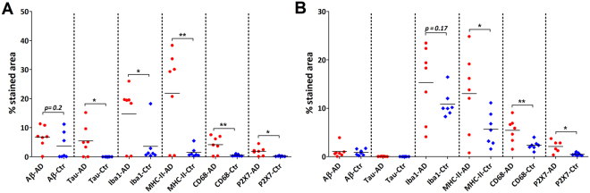
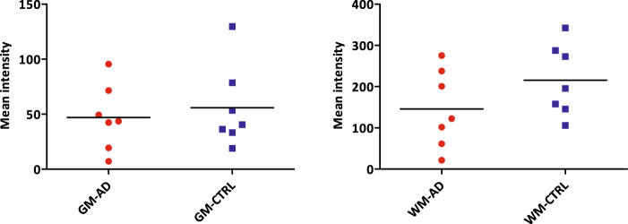
The P2X7 receptor plays a significant role in microglial activation, and as a potential drug target, the P2X7 receptor is also an interesting target in positron emission tomography. The current study aimed at the development and evaluation of a potent tracer targeting the P2X7 receptor, to which end four adamantanyl benzamide analogues with high affinity for the human P2X7 receptor were labelled with carbon-11. All four analogues could be obtained in excellent radiochemical yield and high radiochemical purity and molar activity, and all analogues entered the rat brain. [11C]SMW139 showed the highest metabolic stability in rat plasma, and showed high binding to the hP2X7 receptor in vivo in a hP2X7 receptor overexpressing rat model. Although no significant difference in binding of [11C]SMW139 was observed between post mortem brain tissue of Alzheimer's disease patients and that of healthy controls in in vitro autoradiography experiments, [11C]SMW139 could be a promising tracer for P2X7 receptor imaging using positron emission tomography, due to high receptor binding in vivo in the hP2X7 receptor overexpressing rat model. However, further investigation of both P2X7 receptor expression and binding of [11C]SMW139 in other neurological diseases involving microglial activation is warranted.


Conflict of interest statement

The authors declare no competing interests.

Figures

Figure 1
Figure 1
Synthesis of adamantanyl benzamide precursors 58. Reagents and conditions: (a) BBr3, CH2Cl2, 0 °C to rt, 48 h, quant. (b) PyBOP, DiPEA, CH2Cl2, rt, 18 h, 73% (6), 77% (7), 79% (8).
Figure 2
Figure 2
Carbon-11 methylation of benzamide analogues 13 and [11C]SMW139.
Figure 3
Figure 3
Ex vivo biodistribution following intravenous administration of 13–23 MBq of [11C]1–3 and [11C]SMW139 in healthy male Wistar rats (n = 3 per tracer per time point). Good initial brain uptake was observed for all four analogues. Data are expressed as standardized uptake value (SUV) ± standard error of the mean (SEM).
Figure 4
Figure 4
Autoradiograms of transversal cryosections of rAAV-3flag-hP2X7R vector injected rat brain. (A) Increased [11C]SMW139 binding is observed in rAAV_3flag-hP2X7R injected striatum (left on image). (B) [11C]SMW139 binding in rAAV_3flag-hP2X7R striatum is blocked (79 ± 10%, n = 2) by co-incubation with A-740003 (10 µM). (C) [11C]SMW139 binding in rAAV_3flag-hP2X7R striatum is blocked (93 ± 1%, n = 2) by co-incubation with JNJ-47965567 (10 µM).
Figure 5
Figure 5
Immunohistochemical staining for P2X7R of transversal cryosections of rAAV-3flag-hP2X7R vector injected rat brain. Left: rAAV_3flag-eGFP injected striatum with magnification in upper left corner. Right: rAAV_3flag-hP2X7R injected striatum with magnification in upper right corner.
Figure 6
Figure 6
PET imaging with [11C]SMW139 in rAAV_3flag-hP2X7R rat model. (A) Representative PET-CT image (summed images 0–60 min) of [11C]SMW139 showing increased uptake in rAAV_3flag-hP2X7R injected striatum compared with rAAV_3flag-eGFP (control vector) injected striatum (n = 3). (B) Time-activity curves (TACs) of rAAV_3flag-hP2X7R injected striatum and contralateral (rAAV_3flag-eGFP injected) striatum. Ellipsoid regions of interest (ROIs) were defined within striatum (dimensions: x = 3.4 mm, y = 3.6 mm, z = 2.9 mm) (C) Representative PET-CT image (summed images 0–60 min) of [11C]SMW139 after pre-treatment with JNJ-47965567 (30 mg·kg−1 s.c., 45 min prior to tracer injection). (D) TACs of rAAV_3flag-hP2X7R injected and contralateral striatum after pre-treatment. Ellipsoid ROIs were defined within striatum (dimensions: x = 3.4 mm, y = 3.6 mm, z = 2.9 mm).
Figure 7
Figure 7
Autoradiograms of post mortem human brain tissue (temporal cortex). (A) Total binding of [11C]SMW139 (28 nM). Upper row: AD patients, lower row: non-neurological controls (CTRL). Binding of [11C]SMW139 does not differ significantly between AD patients and controls. Higher tracer binding was observed in white matter compared with grey matter. (B) Binding of [11C]SMW139 could be fully blocked with 10 µM of JNJ-47965567 in both AD patients (upper row) and non-neurological age-matched controls (lower row).
Figure 8
Figure 8
Representative images of immunohistochemical stainings on post mortem brain material of AD patients and non-neurological controls. Overall, staining for all markers is more pronounced in AD patients compared with non-neurological controls (CTRL), particularly in grey matter (GM). Staining for P2X7R is slightly, but significantly increased in both GM and WM of AD patients vs. CTRL. Scale bars indicate 100 µm, except in MHC-II images (50 µm).
Figure 9
Figure 9
Quantification of immunohistochemical staining in human post mortem brain tissue. (A) Quantification of immunohistochemical staining in grey matter depicted per marker (Aβ, tau, Iba1, MHC-II, CD68 and P2X7R). Levels of pathological markers Aβ and tau were higher in AD patients (red circles) than in controls (blue diamonds) Levels of microglial markers Iba1, MHC-II, CD68 and P2X7R were significantly higher in AD patients compared with controls. *p < 0.05, **p < 0.01. (B) Quantification of immunohistochemical staining in white matter depicted per marker (Aβ, tau, Iba1, MHC-II, CD68 and P2X7R). Levels of microglial markers MHC-II, CD68 and P2X7R were significantly higher in AD patients (red circles) compared with controls (blue diamonds). *p < 0.05, **p < 0.01.
Figure 10
Figure 10
Quantification of binding of [11C]SMW139 in autoradiography experiments with human post mortem brain tissue. Both in grey (left) and white (right) matter, no significant differences were observed between AD patients (red circles) and non-neurological controls (blue squares).

References

    1. Beamer E, et al. Purinergic mechanisms in neuroinflammation: An update from molecules to behaviour. Neuropharmacology. 2016;104:94–104. doi: 10.1016/j.neuropharm.2015.09.019. - DOI - PubMed
    1. Bhattacharya A, Biber K. The Microglial ATP-Gated Ion Channel P2X7 As a CNS Drug Target. GLIA. 2016;64:1772–1787. doi: 10.1002/glia.23001. - DOI - PubMed
    1. Burnstock G. P2X ion channel receptors and inflammation. Purinergic Signalling. 2016;12:59–67. doi: 10.1007/s11302-015-9493-0. - DOI - PMC - PubMed
    1. Janssen B, et al. Synthesis and initial preclinical evaluation of the P2X7 receptor antagonist [11C]A-740003 as a novel tracer of neuroinflammation. J. Labelled Compd. Radiopharm. 2014;57:509–516. doi: 10.1002/jlcr.3206. - DOI - PubMed
    1. Gao M, Wang M, Green MA, Hutchins GD, Zheng Q-H. Synthesis of [11C]GSK1482160 as a new PET agent for targeting P2X7 receptor. Bioorg. Med. Chem. Lett. 2016;25:1965–1970. doi: 10.1016/j.bmcl.2015.03.021. - DOI - PubMed

Publication types

MeSH terms

Substances