

CIC-NUTM1 fusion: A case which expands the spectrum of NUT-rearranged epithelioid malignancies
- PMID: 29700887
- PMCID: PMC6881821
- DOI: 10.1002/gcc.3
CIC-NUTM1 fusion: A case which expands the spectrum of NUT-rearranged epithelioid malignancies
Abstract
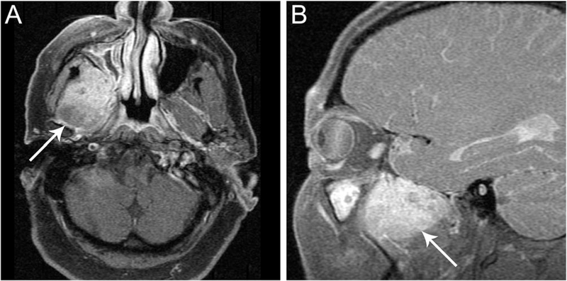
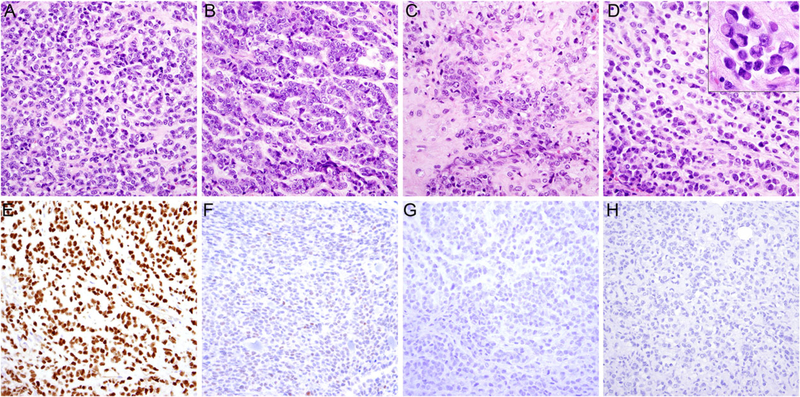
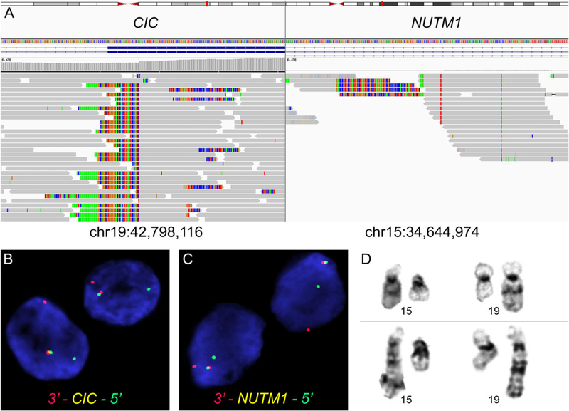
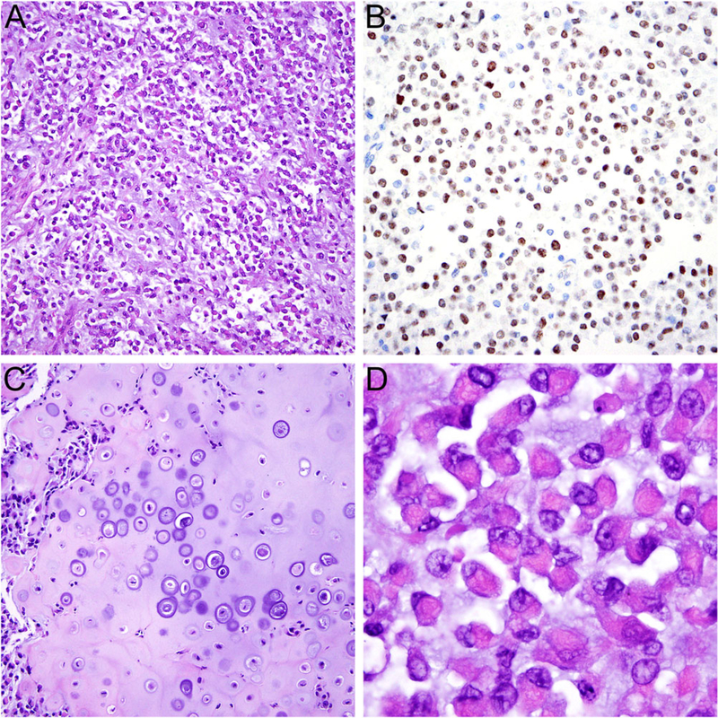
NUT carcinoma (NC) shows very aggressive clinical behavior, occurs predominantly in the thorax and head and neck region of children and adults, and is defined by the presence of NUT (aka NUTM1) rearrangement, mostly BRD4-NUTM1 fusion resulting from t(15;19)(q13; p13.1). So-called "NUT variants" harbor alternate fusions between NUTM1 and BRD3, NSD3, ZNF532, or unknown partners. Rare cases of pediatric tumors with CIC-NUTM1 fusion were recently reported in somatic soft tissue, brain, and kidney. However, such cases have not been identified in adult patients and the presence of a fusion between CIC, characteristic of CIC-rearranged sarcoma, and NUTM1-a defining feature of NC-poses a diagnostic challenge. We herein report a case of malignant epithelioid neoplasm with myoepithelial features harboring CIC-NUTM1 fusion arising in soft tissue of the head in a 60-year-old man. Immunohistochemistry revealed strong expression of NUT, but only weak ETV4 staining and negativity for keratins, EMA, p40, CD99, and WT1. SMARCB1 expression was retained. Fluorescence in situ hybridization and targeted next-generation sequencing identified a CIC-NUTM1 fusion resulting from t(15;19)(q14;q13.2). In light of morphologic features that overlap with those of NC from typical anatomical sites we have seen previously, the tumor was best classified as falling within the NC spectrum rather than CIC-associated sarcoma. This case highlights the emerging diagnostic challenges generated by newly detected gene fusions of unknown clinical and biologic significance. Careful integration of cytogenetic, molecular, and immunohistochemical findings with morphologic appearances in the diagnostic workup of undifferentiated neoplasms is essential.
© 2018 Wiley Periodicals, Inc.
Figures
Similar articles
-
CIC::NUTM1 sarcomas occurred in soft tissues of upper limbs : a rare case report and literature review.Diagn Pathol. 2024 Jun 8;19(1):76. doi: 10.1186/s13000-024-01499-w. Diagn Pathol. 2024. PMID: 38851744 Free PMC article. Review.
-
Primary malignant epithelioid and rhabdoid tumor of bone harboring ZNF532-NUTM1 fusion: the expanding NUT cancer family.Genes Chromosomes Cancer. 2019 Nov;58(11):809-814. doi: 10.1002/gcc.22785. Epub 2019 Jul 30. Genes Chromosomes Cancer. 2019. PMID: 31334571
-
Misleading Germ Cell Phenotype in Pulmonary NUT Carcinoma Harboring the ZNF532-NUTM1 Fusion.Am J Surg Pathol. 2022 Feb 1;46(2):281-288. doi: 10.1097/PAS.0000000000001774. Am J Surg Pathol. 2022. PMID: 34232599 Free PMC article.
-
MAD::NUT Fusion Sarcoma: A Sarcoma Class With NUTM1, NUTM2A, and NUTM2G Fusions and Possibly Distinctive Subtypes.Mod Pathol. 2025 May;38(5):100729. doi: 10.1016/j.modpat.2025.100729. Epub 2025 Feb 6. Mod Pathol. 2025. PMID: 39921028
-
Primary Spindle Cell Sarcoma of the Lung with MGA::NUTM1 Fusion: An Extremely Rare Case of a Potentially Emerging Entity and Review of the Literature.Int J Surg Pathol. 2022 Dec;30(8):931-938. doi: 10.1177/10668969221092125. Epub 2022 Apr 7. Int J Surg Pathol. 2022. PMID: 35388715 Review.
Cited by
-
Update on genetically defined lung neoplasms: NUT carcinoma and thoracic SMARCA4-deficient undifferentiated tumors.Virchows Arch. 2021 Jan;478(1):21-30. doi: 10.1007/s00428-020-03011-3. Epub 2021 Jan 6. Virchows Arch. 2021. PMID: 33409598 Review.
-
NUT Carcinoma: Clinicopathologic Features, Molecular Genetics and Epigenetics.Front Oncol. 2022 Mar 16;12:860830. doi: 10.3389/fonc.2022.860830. eCollection 2022. Front Oncol. 2022. PMID: 35372003 Free PMC article. Review.
-
Salivary Gland NUT Carcinoma with Prolonged Survival in Children: Case Illustration and Systematic Review of Literature.Head Neck Pathol. 2021 Mar;15(1):236-243. doi: 10.1007/s12105-020-01141-3. Epub 2020 Feb 19. Head Neck Pathol. 2021. PMID: 32077054 Free PMC article.
-
The expanding universe of NUTM1 fusions in pediatric cancer.Clin Transl Sci. 2023 Aug;16(8):1331-1339. doi: 10.1111/cts.13535. Epub 2023 May 14. Clin Transl Sci. 2023. PMID: 37082775 Free PMC article. Review.
-
Negative MAPK-ERK regulation sustains CIC-DUX4 oncoprotein expression in undifferentiated sarcoma.Proc Natl Acad Sci U S A. 2020 Aug 25;117(34):20776-20784. doi: 10.1073/pnas.2009137117. Epub 2020 Aug 11. Proc Natl Acad Sci U S A. 2020. PMID: 32788348 Free PMC article.
References
-
- French CA,Miyoshi I,Kubonishi I,Grier HE,Perez-Atayde AR, Fletcher JA. 2003. BRD4-NUT fusion oncogene: a novel mechanism in aggressive carcinoma. Cancer Res. 63:304–307. - PubMed
-
- French CA,Ramirez CL,Kolmakova J, et al. 2008. BRD-NUT oncoproteins: a family of closely related nuclear proteins that block epithelial differentiation and maintain the growth of carcinoma cells. Oncogene. 27:2237–2242. - PubMed
Publication types
MeSH terms
Substances
Grants and funding
LinkOut - more resources
Full Text Sources
Other Literature Sources
Medical
Research Materials
Miscellaneous
