

FaptaSyme: A Strategy for Converting a Monomer/Oligomer-Nonselective Aptameric Sensor into an Oligomer-Selective One
- PMID: 29700982
- PMCID: PMC6422747
- DOI: 10.1002/cbic.201800017
FaptaSyme: A Strategy for Converting a Monomer/Oligomer-Nonselective Aptameric Sensor into an Oligomer-Selective One
Abstract
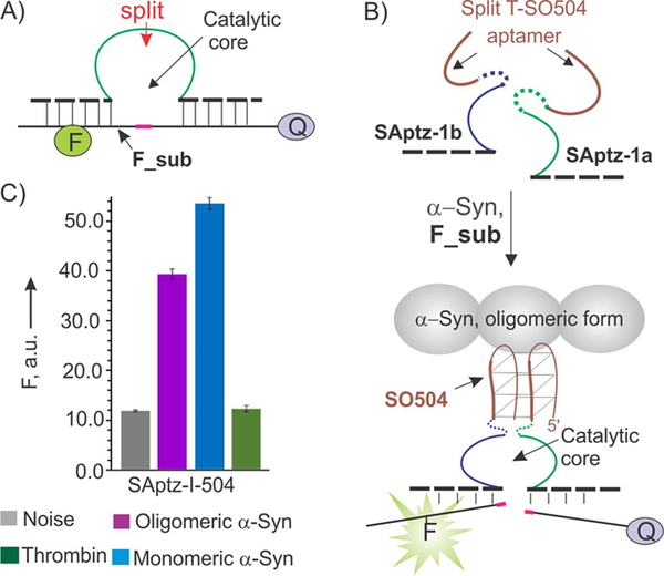
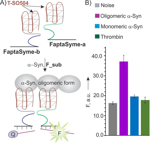
Aptameric sensors can bind molecular targets and produce output signals, a phenomenon that is used in bioassays. In some cases, it is important to distinguish between monomeric and oligomeric forms of a target. Here, we propose a strategy to convert a monomer/oligomer-nonselective sensor into an oligomer-selective sensor. We designed an aptazyme that produced a high fluorescent output in the presence of oligomeric α-synuclein (a molecular marker of Parkinson's disease) but not its monomeric form. The strategy is potentially useful in the design of point-of-care tests for the diagnosis of neurodegenerative diseases.
Keywords: aptamers; aptazymes; biosensors; deoxyribozymes; split probes.
© 2018 Wiley-VCH Verlag GmbH & Co. KGaA, Weinheim.
Figures
References
-
- Ellington AD and Szostak JW, Nature 1990, 346, 818–22; - PubMed
- Mercier MC, Dontenwill M and Choulier L, Cancers (Basel) 2017, 9; - PMC - PubMed
- Sefah K, Phillips JA, Xiong X, Meng L, Van Simaeys D, Chen H, Martin J and Tan W, Analyst 2009, 134, 1765–1775; - PubMed
- Pfeiffer F, Rosenthal M, Siegl J, Ewers J and Mayer G, Curr Opin Biotechnol 2017, 48, 111–118; - PubMed
- Reverdatto S, Burz DS and Shekhtman A, Curr Top Med Chem 2015, 15, 1082–1101. - PMC - PubMed
-
- Thean D, Ebo JS, Luxton T, Lee XC, Yuen TY, Ferrer FJ, Johannes CW, Lane DP and Brown CJ, Sci Rep 2017, 7, 1763; - PMC - PubMed
- Ding F, Gao Y and He X, Bioorg Med Chem Lett 2017, 27, 4256–4269; - PubMed
- Farzin L, Shamsipur M and Sheibani S, Talanta 2017, 174, 619–627; - PubMed
- Alizadeh N, Memar MY, Moaddab SR and Kafil HS, Biomed. Pharmacother 2017, 93, 737–745; - PubMed
- Tang J, Huang N, Zhang X, Zhou T, Tan Y, Pi J, Pi L, Cheng S, Zheng H and Cheng Y, Int. J. Nanomedicine 2017, 12, 3899–3911; - PMC - PubMed
- Cho EJ, Lee JW and Ellington AD, Annu. Rev. Anal. Chem. (Palo Alto Calif) 2009, 2, 241–264; - PubMed
- Labib M and Berezovski MV, Adv. Biochem. Eng. Biotechnol 2014, 140, 155–181. - PubMed
-
- Yamamoto R, Baba T and Kumar PK, Genes Cells 2000, 5, 389–396; - PubMed
- Yamamoto-Fujita R and Kumar PK, Anal. Chem 2005, 77, 5460–5466; - PubMed
- Yoshida W, Sode K and Ikebukuro K, Biotechnol. Lett 2008, 30, 421–425; - PubMed
- Xu W and Lu Y, Anal Chem 2010, 82, 574–578. - PMC - PubMed
- Sosic A, Meneghello A, Cretaio E and Gatto B, Sensors (Basel) 2011, 11, 9426–9441; - PMC - PubMed
- Lin Z, Chen L, Zhu X, Qiu B and Chen G, Chem Commun (Camb) 2010, 46, 5563–5565; - PubMed
- Yu H, Canoura J, Guntupalli B, Lou X and Xiao Y, Chem. Sci 2017, 8, 131–141; - PMC - PubMed
- Yuan B, Zhou Y, Guo Q, Wang K, Yang X, Meng X, Wan J, Tan Y, Huang Z, Xie Q and Zhao X, Chem. Commun 2016, 52, 1590–1593. - PubMed
Grants and funding
LinkOut - more resources
Full Text Sources
Other Literature Sources
