. 2018 Jul:17:171-179.
doi: 10.1016/j.redox.2018.04.013. Epub 2018 Apr 16.

MGST1, a GSH transferase/peroxidase essential for development and hematopoietic stem cell differentiation

Affiliations

MGST1, a GSH transferase/peroxidase essential for development and hematopoietic stem cell differentiation

Lars Bräutigam et al. Redox Biol. 2018 Jul.

Abstract

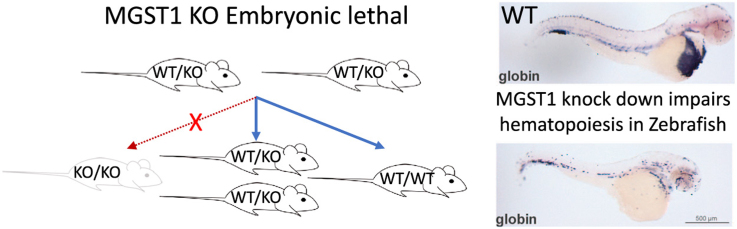
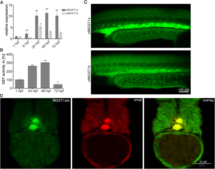
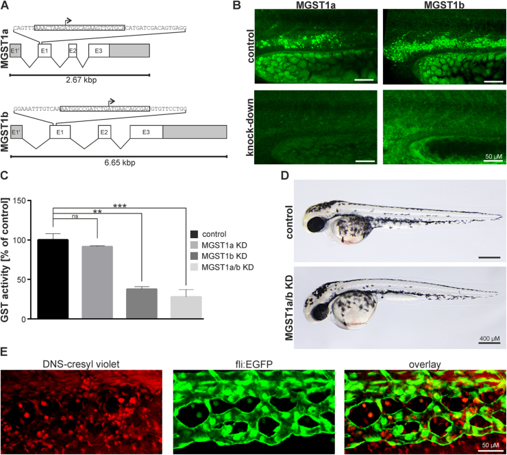
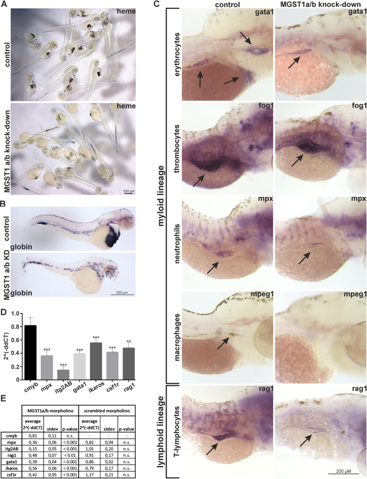
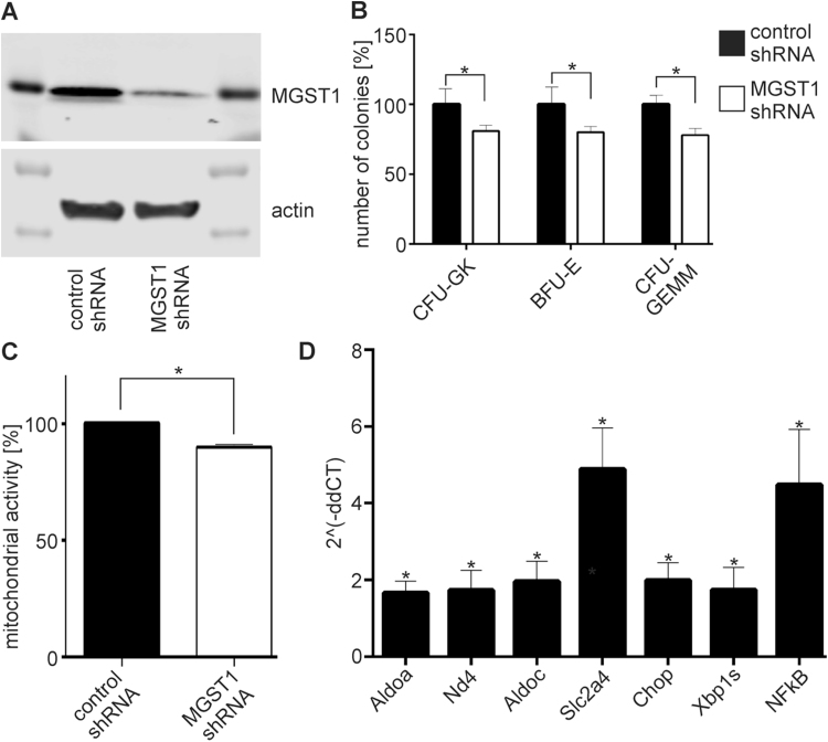
We show for the first time that, in contrast to other glutathione transferases and peroxidases, deletion of microsomal glutathione transferase 1 (MGST1) in mice is embryonic lethal. To elucidate why, we used zebrafish development as a model system and found that knockdown of MGST1 produced impaired hematopoiesis. We show that MGST1 is expressed early during zebrafish development and plays an important role in hematopoiesis. High expression of MGST1 was detected in regions of active hematopoiesis and co-expressed with markers for hematopoietic stem cells. Further, morpholino-mediated knock-down of MGST1 led to a significant reduction of differentiated hematopoietic cells both from the myeloid and the lymphoid lineages. In fact, hemoglobin was virtually absent in the knock-down fish as revealed by diaminofluorene staining. The impact of MGST1 on hematopoiesis was also shown in hematopoietic stem/progenitor cells (HSPC) isolated from mice, where it was expressed at high levels. Upon promoting HSPC differentiation, lentiviral shRNA MGST1 knockdown significantly reduced differentiated, dedicated cells of the hematopoietic system. Further, MGST1 knockdown resulted in a significant lowering of mitochondrial metabolism and an induction of glycolytic enzymes, energetic states closely coupled to HSPC dynamics. Thus, the non-selenium, glutathione dependent redox regulatory enzyme MGST1 is crucial for embryonic development and for hematopoiesis in vertebrates.

Keywords: Embryonic development; Hematopoiesis; Microsomal glutathione transferase/peroxidase; Redox regulation.


Figures

fx1
Graphical abstract
Fig. 1
Fig. 1
Expression of MGST1 during zebrafish embryo development. (A) Relative expression of zfMGST1a/b during zebrafish embryonic development. (B) Relative activity of GST enzymes during zebrafish embryonic development. (C) Expression of MGST1a and MGST1b in the intermediate cell mass of 24 hpf embryos. (D) MGST1a/b protein colocalizes with cmyb, a marker specific for HSCs, in 48 hpf embryos. Confocal images were taken on a Leica LSM700 with a 20× lens; Alexa 488 and 555 filters were used; images stacks were produced with ImageJ, GIMP was used to adjust the gamma channel.
Fig. 2
Fig. 2
Morpholino induced knock-down of MGST1 in zebrafish. (A) Genomic organization of zfMGST1 and location of morpholino attachment sites targeting transcription. (B) Immunohistochemistry in embryos injected with morpholinos knocking down MGST1a/b. (C) Enzymatic activity of GST enzymes in extracts of embryos injected with morpholinos knocking-down MGST1a, MGST1b or both. (D) Gross morphology of embryos injected with morpholinos knocking-down MGST1a/b. (E) DNS-cresyl violet staining indicating MGST1 positive cells in the caudal hematopoietic tissue, see also Supplementary movie S1. Brightfield images were taken on a Leica MZ16 microscope equipped with a Leica DFC300FX camera; Confocal images were taken on a Leica LSM700 with a 20× lens; Alexa 488 and 555 filters were used; images stacks were produced with ImageJ, GIMP was used to adjust the gamma channel.
Movie S1
Movie S1
48 h old zebrafish embryos were mounted in low melting agarose and incubated with 2.5 µM fluorogenic GST substrate 2,4-dinitrobenzene sulfonamide cresyl violet. Images were recorded with excitation at 540 nm and emission at 620 nm. ISV: intersegmental vessels; DA: dorsal aorta; CV: caudal vein. Arrow follows GST stained and mobile cell.
Fig. 3
Fig. 3
Loss of functional MGST1 blocks differentiation of hematopoietic stem cells in zebrafish. (A) Histochemical staining of hemoglobin in living zebrafish embryos. (B) Whole-mount in situ hybridization in 48 hpf embryos staining globin transcripts specific for erythrocytes. (C) Whole-mount in situ hybridization against marker genes for different myloid and lymphoid lineages in 96 hpf embryos. (D) RTqPCR quantifying transcripts specific for different myloid and lymphoid lineages in 96 hpf embryos. (E) Quantification and statistics for RTqPCR. Brightfield images were taken on a Leica MZ16 microscope equipped with a Leica DFC300FX camera; images stacks were produced with ImageJ, GIMP was used to adjust the gamma channel.
Fig. 4
Fig. 4
Loss of functional MGST1 blocks differentiation of HSPC cells and induces metabolic changes. (A) HSPC cells were transfected with a lentiviral-scrambled (control) shRNA or a lentiviral-shRNA against MGST1. Immunoblot analysis indicated that shRNA against MGST1 significantly reduced the protein levels in HSPC. (B) MGST1 knock-down in HSPC resulted in decreased differentiation. Colony forming unit (CFU) assays were used and CFU-granulocyte macrophage (GM), burst forming unit erythroid (BFU-E) and CFU- granulocyte, erythrocyte, monocyte and megakaryocyte (GEMM) colonies were counted. (C) Mitochondrial activity was assessed with the MTT assay. (D) Quantitative RT-PCR analysis of the expression of key genes associated with glycolysis, HIF1 controlled genes and NFkB. Data are presented as MGST1 knock-down HSPC relative to controls. Data are means (+/− SD) for at least three experiments *p < 0.05.

References

    1. Hayes J.D., Flanagan J.U., Jowsey I.R. Glutathione transferases. Annu. Rev. Pharmacol. Toxicol. 2005;45:51–88. - PubMed
    1. Rinaldi R., Eliasson E., Swedmark S., Morgenstern R. Reactive intermediates and the dynamics of glutathione transferases. Drug Metab. Dispos. 2002;30(10):1053–1058. - PubMed
    1. Sies H., Berndt C., Jones D.P. Oxidative stress. Annu. Rev. Biochem. 2017;86:715–748. - PubMed
    1. Arner E.S. Focus on mammalian thioredoxin reductases–important selenoproteins with versatile functions. Biochim. Biophys. Acta. 2009;1790(6):495–526. - PubMed
    1. Brigelius-Flohe R., Maiorino M. Glutathione peroxidases. Biochim. Biophys. Acta. 2013;1830(5):3289–3303. - PubMed

Publication types

MeSH terms