Review
. 2018 Apr 27;19(5):1306.
doi: 10.3390/ijms19051306.

Positive and Negative Regulation of Angiogenesis by Soluble Vascular Endothelial Growth Factor Receptor-1

Affiliations
Review

Positive and Negative Regulation of Angiogenesis by Soluble Vascular Endothelial Growth Factor Receptor-1

Cristina M Failla et al. Int J Mol Sci. .

Abstract

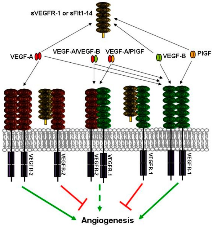
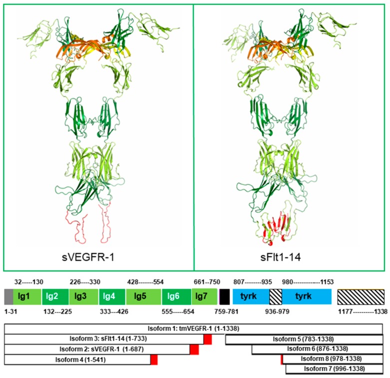
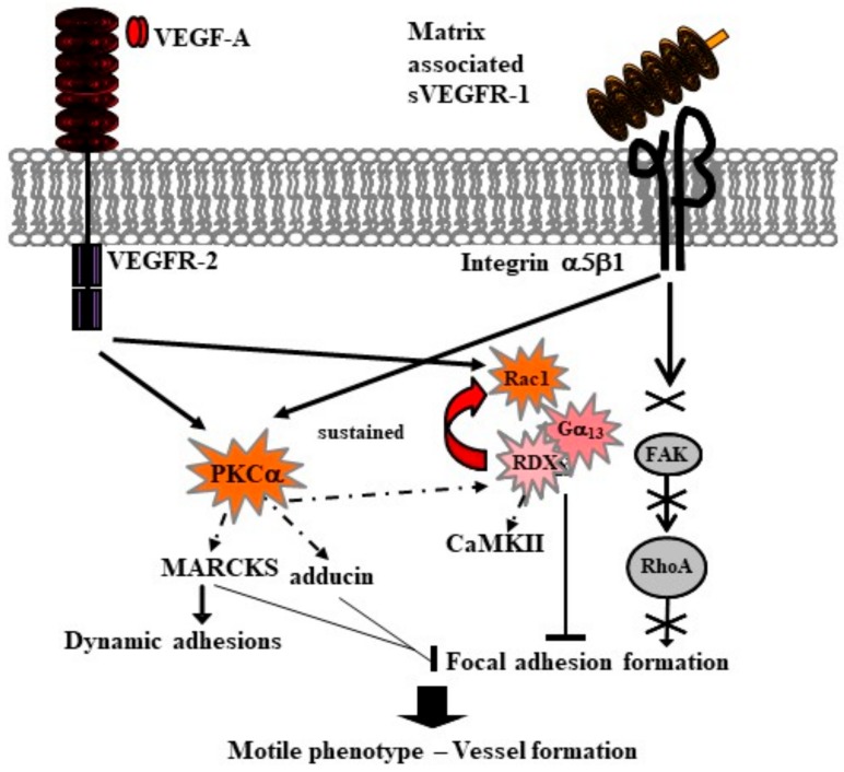
Vascular endothelial growth factor receptor (VEGFR)-1 exists in different forms, derived from alternative splicing of the same gene. In addition to the transmembrane form, endothelial cells produce a soluble VEGFR-1 (sVEGFR-1) isoform, whereas non-endothelial cells produce both sVEGFR-1 and a different soluble molecule, known as soluble fms-like tyrosine kinase (sFlt)1-14. By binding members of the vascular endothelial growth factor (VEGF) family, the soluble forms reduce the amounts of VEGFs available for the interaction with their transmembrane receptors, thereby negatively regulating VEGFR-mediated signaling. In agreement with this activity, high levels of circulating sVEGFR-1 or sFlt1-14 are associated with different pathological conditions involving vascular dysfunction. Moreover, sVEGFR-1 and sFlt1-14 have an additional role in angiogenesis: they are deposited in the endothelial cell and pericyte extracellular matrix, and interact with cell membrane components. Interaction of sVEGFR-1 with α5β1 integrin on endothelial cell membranes regulates vessel growth, triggering a dynamic, pro-angiogenic phenotype. Interaction of sVEGFR-1/sFlt1-14 with cell membrane glycosphingolipids in lipid rafts controls kidney cell morphology and glomerular barrier functions. These cell⁻matrix contacts represent attractive novel targets for pharmacological intervention in addition to those addressing interactions between VEGFs and their receptors.

Keywords: Vascular endothelial growth factor receptor; angiogenesis; extracellular matrix.


Conflict of interest statement

The authors declare no conflict of interest.

Figures

Figure 1
Figure 1
Anti-angiogenic roles of soluble vascular endothelial growth factor receptor (VEGFR)-1 VEGFR-1 isoforms. Soluble VEGFR-1 isoforms, either sVEGFR-1 or soluble fms-like tyrosine kinase (sFlt)1-14, act as “VEGF sinks”. By binding members of the VEGF family, they decrease the amount of growth factors available to interact with the transmembrane tyrosine kinase receptors. In addition, soluble receptor isoforms can reduce VEGF signal transduction by binding to transmembrane receptor monomers and blocking formation of signaling competent receptor homodimers. Receptor heterodimers can also be inhibited by soluble isoforms, thus blocking the signaling of VEGF-A/placenta growth factor (PlGF) or VEGF-A/VEGF-B heterodimers. Interactions that lead to and hamper angiogenesis are indicated by green and red arrows/lines, respectively. The dashed arrow indicates the limited pro-angiogenic activity of receptor heterodimers.
Figure 2
Figure 2
VEGFR-1 structures. Top: Soluble isoforms sVEGFR-1 (left) and sFlt1-14 (right) are shown by ribbon representation. The 3D structure of Ig-like domains 1–6 (residues 1–654), common to both isoforms, has been experimentally determined in complex with VEGF-A (PDB ID: 5T89, Resolution: 4 Å). VEGF monomers are orange and yellow. VEGFR-1 Ig-like domains 1, 3 and 5 are light green; Ig-like domains 2, 4 and 6 are dark green; residues 657–705 of sFlt-1, whose sequence is identical to transmembrane VEGFR-1 (isoform 1) are light green; the 30 residues C-terminal tail of sVEGFR-1 (657–687) and residues 706–733 of sFlt-1, whose sequence differs from isoform 1 are red. The C-terminal regions of sVEGFR-1 and sFlt1-14 have been modeled by homology using the structure of VEGFR-2 domain 7 (PDB ID: 3KVQ, Resolution: 2.7 Å) as a template, using the molecular graphics program InsightII (Accelrys Software Inc., [26]). However, both these regions are likely to be, at least in the red-colored part, disordered (see text). Bottom: Domain architecture of VEGFR-1 isoforms. VEGFR-1 is color coded as follows: N-terminal signal sequence, grey; Ig-like domains 1, 3, 5 and 7, light green; Ig-like domains 2, 4, and 6, dark green; transmembrane helix, black; tyrosine kinase domain, blue; disordered regions, striped. Isoform sequences that differ from transmembrane VEGFR-1 are red. Boundaries of Ig-like domains 1–6 are defined according to the experimental structure. All the other regions are defined based on a combination of programs for domain assignment (i.e., SMART [27], Pfam [28], Superfamily [29]) and transmembrane helices (i.e., TMHMM [30]) or secondary structure and disordered region predictions (i.e., PsiPred [25]). The length of each segment in the picture is proportional to the number of residues comprised in each domain or region.
Figure 3
Figure 3
Molecular signals of motility are activated by endothelial cell adhesion to matrix sVEGFR-1 through α5β1 integrin. Protein kinase C alpha (PKCα) phosphorylation by VEGFR-2 and/or α5β1 integrin (arrows) modulates both expression and phosphorylation (dotted arrow) of its substrates adducin, myristoylated alanine-rich protein kinase C substrate (MARCKS), and radixin (RDX) (see text). VEGFR-2 starts a signaling loop that involves the Rho family GTPase Rac1 and the heterotrimeric G protein α13 (Gα13), and is sustained (red arrow) by the activation of radixin, which also phosphorylates the calcium/calmodulin-dependent protein kinase II (CaMKII). Cell adhesion to matrix sVEGFR-1 does not activate the focal adhesion kinase (FAK) signaling (cross and crossed arrows). As a consequence, focal adhesion formation is blocked (bars). MARCKS expression is increased, and non-phosphorylated MARCKS accumulates into and stabilizes dynamic adhesions (black arrow). Thus, endothelial cells acquire a highly motile phenotype that allows them to initiate new vessel formation (thick black arrow).

References

    1. Risau W. Mechanisms of angiogenesis. Nature. 1997;386:671–674. doi: 10.1038/386671a0. - DOI - PubMed
    1. Folkman J. Angiogenesis in cancer, vascular, rheumatoid and other disease. Nat. Med. 1995;1:27–31. doi: 10.1038/nm0195-27. - DOI - PubMed
    1. Shibuya M., Yamaguchi S., Yamane A., Ikeda T., Tojo A., Matsushime H., Sato M. Nucleotide sequence and expression of a novel human receptor-type tyrosine kinase (Flt) closely related to the FMS family. Oncogene. 1990;8:519–527. - PubMed
    1. Cébe-Suarez S., Zehnder-Fjallman A., Ballmer-Hofer K. The role of VEGF receptors in angiogenesis; complex partnerships. Cell. Mol. Life Sci. 2006;63:601–615. doi: 10.1007/s00018-005-5426-3. - DOI - PMC - PubMed
    1. De Vries C., Escobedo J., Ueno H., Houck K., Ferrara N., Williams L. The fms-like tyrosine kinase, a receptor for vascular endothelial growth factor. Science. 1992;255:989–991. doi: 10.1126/science.1312256. - DOI - PubMed

MeSH terms

Substances