. 2018 Sep;65(2):e12501.
doi: 10.1111/jpi.12501. Epub 2018 May 21.

Melatonin and its derivatives counteract the ultraviolet B radiation-induced damage in human and porcine skin ex vivo

Affiliations

Melatonin and its derivatives counteract the ultraviolet B radiation-induced damage in human and porcine skin ex vivo

Cezary Skobowiat et al. J Pineal Res. 2018 Sep.

Expression of concern in

Abstract

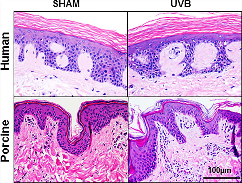
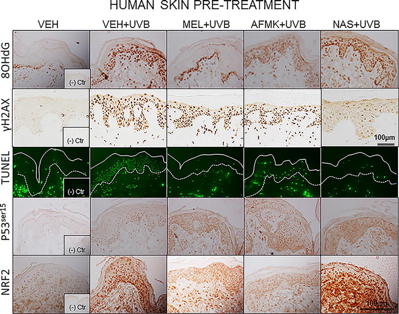
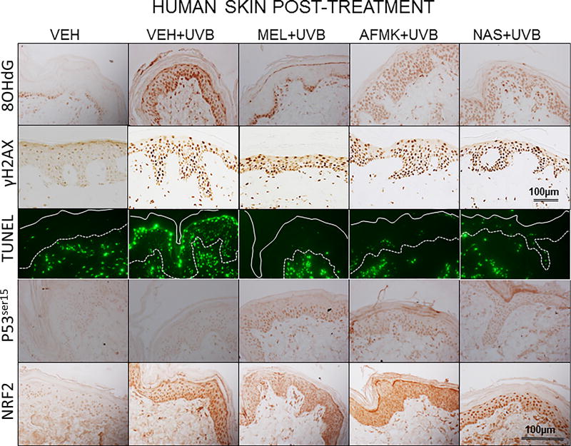
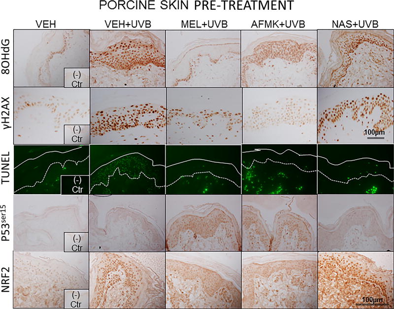
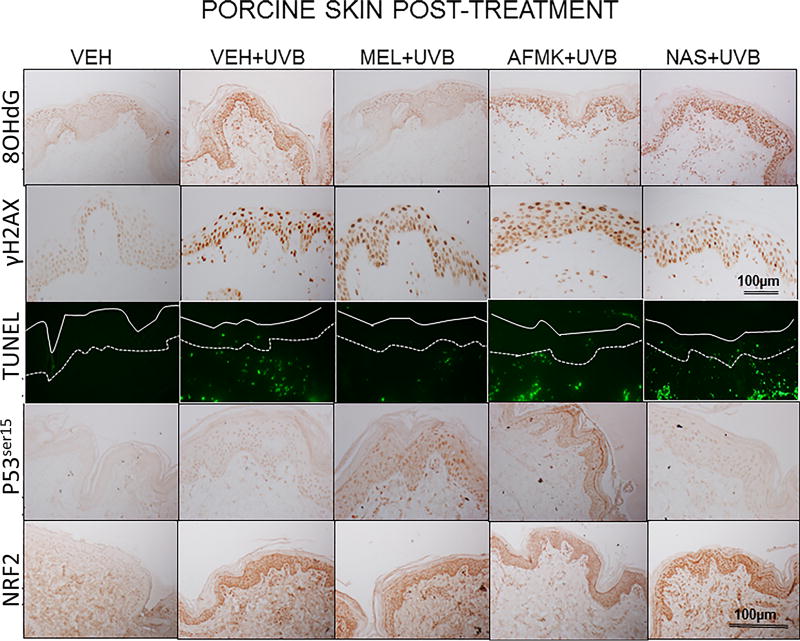
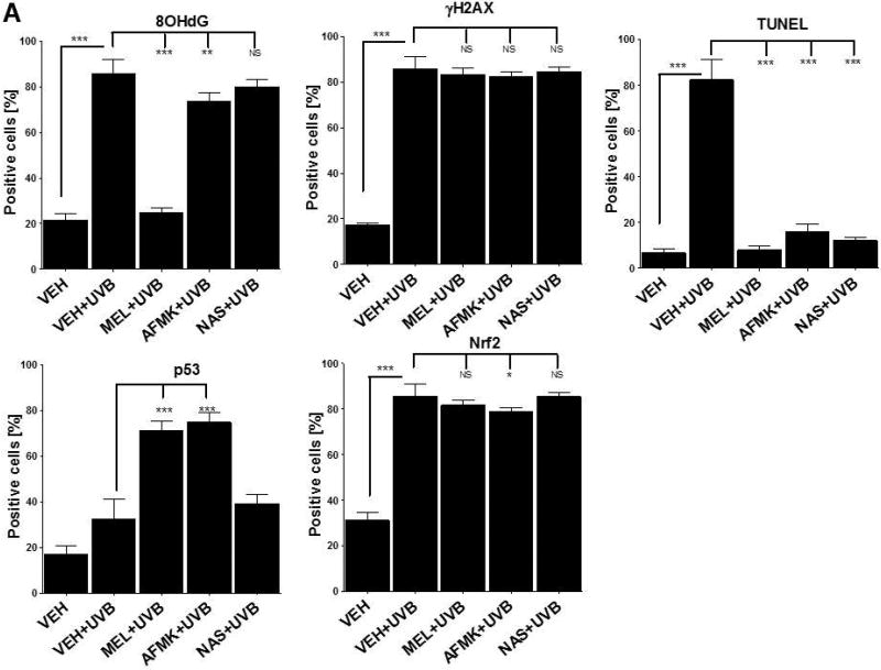
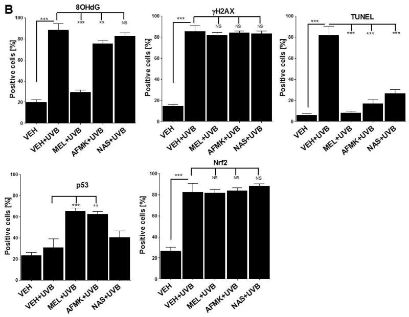
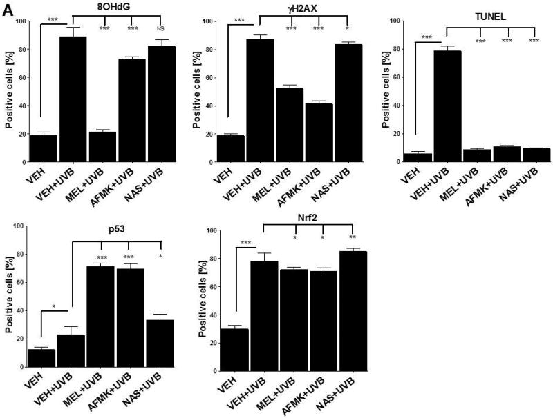
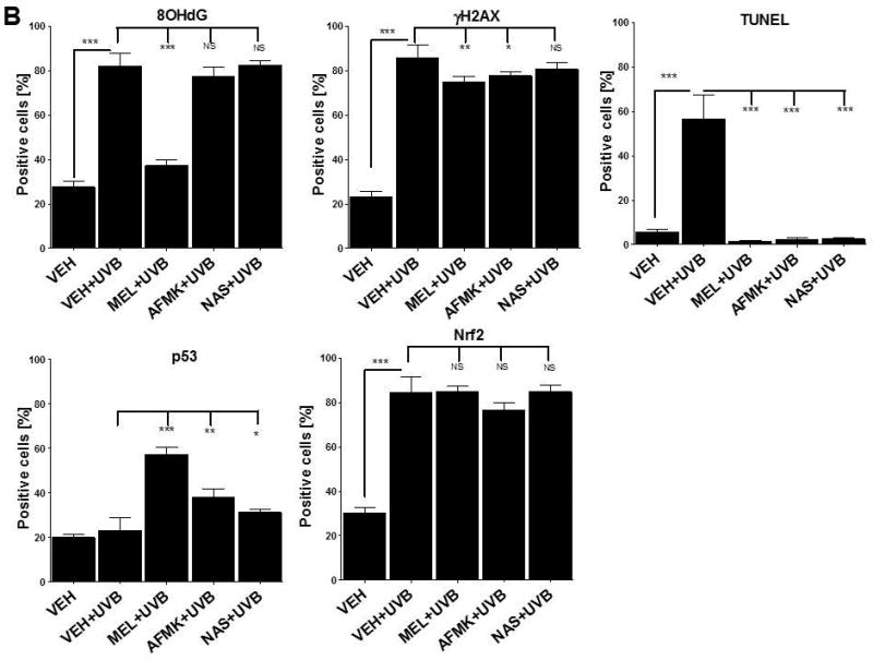
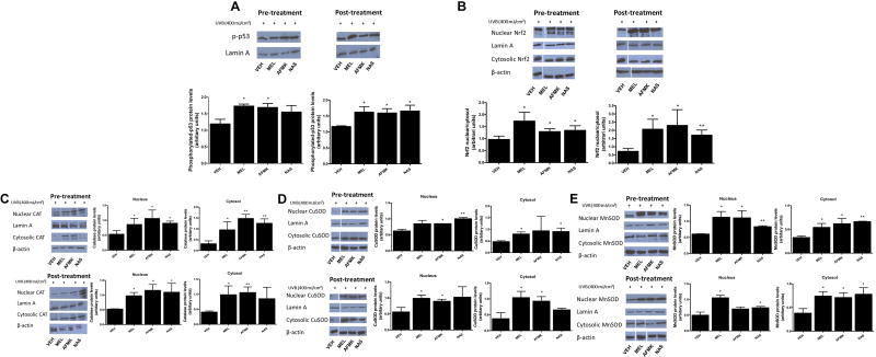
Melatonin and its derivatives (N1 -acetyl-N2 -formyl-5-methoxykynurenine [AFMK] and N-acetyl serotonin [NAS]) have broad-spectrum protective effects against photocarcinogenesis, including both direct and indirect antioxidative actions, regulation of apoptosis and DNA damage repair; these data were primarily derived from in vitro models. This study evaluates possible beneficial effects of melatonin and its active derivatives against ultraviolet B (UVB)-induced harm to human and porcine skin ex vivo and to cultured HaCaT cells. The topical application of melatonin, AFMK, or NAS protected epidermal cells against UVB-induced 8-OHdG formation and apoptosis with a further increase in p53ser15 expression, especially after application of melatonin or AFMK but not after NAS use. The photoprotective action was observed in pre- and post-UVB treatment in both human and porcine models. Melatonin along with its derivatives upregulated also the expression of antioxidative enzymes after UVB radiation of HaCaT cells. The exogenous application of melatonin or its derivatives represents a potent and promising tool for preventing UVB-induced oxidative stress and DNA damage. This protection results in improved genomic, cellular, and tissue integrity against UVB-induced carcinogenesis, especially when applied prior to UV exposure. In addition, our ex vivo experiments provide fundamental justification for further testing the clinical utility of melatonin and metabolites as protectors again UVB in human subjects. Our ex vivo data constitute the bridge between vitro to vivo translation and thus justifies the pursue for further clinical utility of melatonin in maintaining skin homeostasis.

Keywords: UVB; epidermis; melatonin; photoprotection; skin.


Figures

Figure 1
Figure 1
A single (400 mJ/cm2) dose of ultraviolet B (UVB) does not lead to a visible damage of human and porcine skin. There is lack of detectable epidermal necrosis, keratinocyte apoptosis or acute inflammatory responses in UVB-treated and sham-irradiated skin. H&E staining. Scale bar 100µm.
Figure 2
Figure 2
The representative images of the indicators of oxidative stress (8OHdG), DNA damage (γH2AX), apoptosis (TUNEL), and DNA-repair and antioxidative mechanisms (p53ser115 and Nrf2) after a single dose of 400 mJ/cm2 of UVB applied to human skin ex vivo. Skin was topically treated with melatonin, AFMK or NAS 4 h prior to (A) or immediately after irradiation (B).
Figure 2
Figure 2
The representative images of the indicators of oxidative stress (8OHdG), DNA damage (γH2AX), apoptosis (TUNEL), and DNA-repair and antioxidative mechanisms (p53ser115 and Nrf2) after a single dose of 400 mJ/cm2 of UVB applied to human skin ex vivo. Skin was topically treated with melatonin, AFMK or NAS 4 h prior to (A) or immediately after irradiation (B).
Figure 3
Figure 3
The representative images of the indicators of oxidative stress (8OHdG), DNA damage (γH2AX), apoptosis (TUNEL), DNA-repair and antioxidative mechanisms (p53ser115 and Nrf2) after a single dose of 400 mJ/cm2 of UVB applied to porcine skin ex vivo. Skin was topically treated with melatonin, AFMK or NAS 4 h prior to (A) or immediately after irradiation (B).
Figure 3
Figure 3
The representative images of the indicators of oxidative stress (8OHdG), DNA damage (γH2AX), apoptosis (TUNEL), DNA-repair and antioxidative mechanisms (p53ser115 and Nrf2) after a single dose of 400 mJ/cm2 of UVB applied to porcine skin ex vivo. Skin was topically treated with melatonin, AFMK or NAS 4 h prior to (A) or immediately after irradiation (B).
Figure 4
Figure 4
The quantitative analysis of the modulatory effects of topical treatment with melatonin, AFMK or NAS on 8OHdG, γH2AX, TUNEL, p53ser115 and Nrf2 expression in human skin. The compounds were topically applied 4 h prior to (A) or immediately after (B) UVB exposure. Data are presented as the percentage of positively-stained cells per 100 consecutive epidermal cells ± SD; the statistical significance of the differences was evaluated by Student’s t test; where *P < 0.05, **P < 0.01, ***P < 0.0001, and NS – lack of significance.
Figure 4
Figure 4
The quantitative analysis of the modulatory effects of topical treatment with melatonin, AFMK or NAS on 8OHdG, γH2AX, TUNEL, p53ser115 and Nrf2 expression in human skin. The compounds were topically applied 4 h prior to (A) or immediately after (B) UVB exposure. Data are presented as the percentage of positively-stained cells per 100 consecutive epidermal cells ± SD; the statistical significance of the differences was evaluated by Student’s t test; where *P < 0.05, **P < 0.01, ***P < 0.0001, and NS – lack of significance.
Figure 5
Figure 5
The quantitative analysis of the inhibitory effects of topical treatment with melatonin, AFMK or NAS on 8OHdG, γH2AX, TUNEL, p53ser115 and Nrf2 expression in porcine skin. The compounds were topically applied 4 h prior to (A) and immediately after (B) UVB radiation. Data are presented as the percentage of positively-stained cells per 100 consecutive epidermal cells ± SD; the statistical significance of the differences was evaluated by Student’s t test; where *P < 0.05, **P < 0.01, ***P < 0.0001, and NS – lack of statistical significance.
Figure 5
Figure 5
The quantitative analysis of the inhibitory effects of topical treatment with melatonin, AFMK or NAS on 8OHdG, γH2AX, TUNEL, p53ser115 and Nrf2 expression in porcine skin. The compounds were topically applied 4 h prior to (A) and immediately after (B) UVB radiation. Data are presented as the percentage of positively-stained cells per 100 consecutive epidermal cells ± SD; the statistical significance of the differences was evaluated by Student’s t test; where *P < 0.05, **P < 0.01, ***P < 0.0001, and NS – lack of statistical significance.
Figure 6
Figure 6
Treatment with melatonin, AFMK or NAS increased the UVB-induced phosphorylation of p53 in HaCaT cells (A), nuclear translocation of Nrf2 (B), catalase (C), CuSOD (D), and MnSOD (E) protein expression. In pre-treatment experiment, cells were treated with melatonin, AFMK or NAS, at the concentration 5 ×10⁻⁵ M or 0.1% EtOH (vehicle control) for 30 min and subsequently, under PBS, irradiated with UVB 400 mJ/cm2. For post-treatment experiment, cells were irradiated with UVB 400 mJ/cm2 and then treated with melatonin, AFMK or NAS at the concentration 5 ×10⁻⁵ M or 0.1% EtOH (vehicle control) for 30 min. Cells were harvested immediately after treatment and western blotting was performed to evaluate the phosphorylation rate of p53, nuclear translocation of Nrf2, catalase, CuSOD, and MnSOD protein expression. Phosphorylated p53 was detected at 53 kDa, Nrf2 at 61 kDa, catalase at 64 kDa, CuSOD at 23 kDa, MnSOD at 24 kDa, lamin A (a loading control for nuclear protein) at 69 kDa, and β-actin (a loading control for cytosol protein) at 42 kDa. Data was expressed as means ± SD. The statistical significance of differences was evaluated by Student’s t test. *P < 0.05; **P < 0.01 versus EtOH (vehicle control).

References

    1. Brozyna A, Zbytek B, Granese J, et al. Mechanism of UV-related carcinogenesis and its contribution to nevi/melanoma. Expert Rev Dermatol. 2007;2:451–69. - PMC - PubMed
    1. Cadet J, Douki T, RAVANAT JL. Oxidatively generated damage to cellular DNA by UVB and UVA radiation. Photochem Photobiol. 2015;91:140–55. - PubMed
    1. Slominski AT, Zmijewski MA, Skobowiat C, et al. Sensing the environment: regulation of local and global homeostasis by the skin's neuroendocrine system. Adv Anat Embryol Cell Biol. 2012;212:v–vii. 1–115. - PMC - PubMed
    1. Slominski A, Wortsman J. Neuroendocrinology of the skin. Endocr Rev. 2000;21:457–87. - PubMed
    1. Geyer RK, Nagasawa H, Little JB, et al. Role and regulation of p53 during an ultraviolet radiation-induced G1 cell cycle arrest. Cell Growth Differ. 2000;11:149–56. - PubMed