. 2018 Apr 27;15(1):126.
doi: 10.1186/s12974-018-1160-2.

Human immunodeficiency virus type 1 (HIV-1)-mediated neuroinflammation dysregulates neurogranin and induces synaptodendritic injury

Affiliations

Human immunodeficiency virus type 1 (HIV-1)-mediated neuroinflammation dysregulates neurogranin and induces synaptodendritic injury

Debjani Guha et al. J Neuroinflammation. .

Abstract

Background: Human immunodeficiency virus type 1 (HIV-1)-associated neurocognitive disorder (HAND) is a common outcome of a majority of HIV-1-infected subjects and is associated with synaptodendritic damage. Neurogranin (Ng), a postsynaptic protein, and calmodulin (CaM) are two important players of synaptic integrity/functions. The biological role of Ng in the context of HAND is unknown.

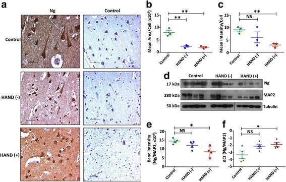
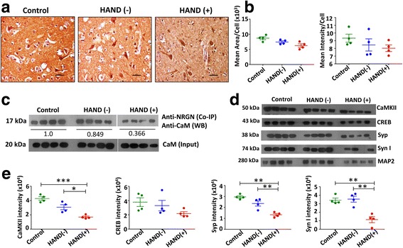
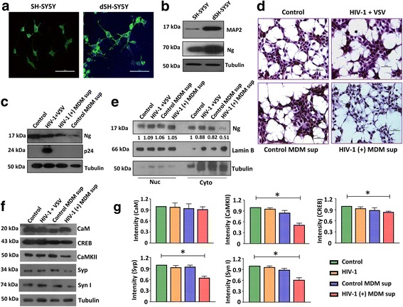
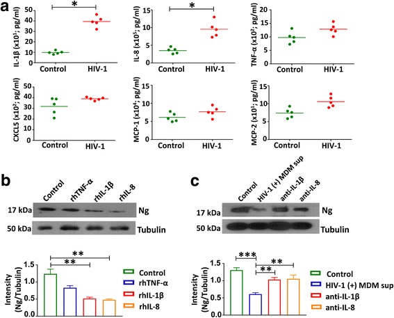
Methods: We compared the expression of Ng in frontal cortex (FC) tissues from control and HIV-1-positive subjects with and without HAND by immunohistochemistry, western blot, and qRT-PCR. The interaction between Ng and CaM was analyzed by co-immunoprecipitation. Ng, microtubule-associated protein 2 (MAP2), CaM, CaM-dependent protein kinase II (CaMKII), CREB, synaptophysin (Syp), and synapsin I (Syn I) expressions were evaluated by western blot using FC tissue lysates and differentiated SH-SY5Y (dSH-SY5Y) cells. Identification of inflammatory factors related to Ng loss was accomplished by exposing dSH-SY5Y cells to HIV-1 and mock-infected monocyte-derived macrophage (MDM) supernatants or HIV-1 NLYU2 pseudotyped with VSV-G-Env. Levels of interleukin (IL)-1β, IL-8, tumor necrosis factor (TNF)-α, monocyte chemoattractant protein (MCP)-1, MCP-2, and CXCL5 in MDM supernatants were measured by ELISA. Association of IL-1β and IL-8 to Ng expression in context of HIV-1 infection was evaluated in the presence or absence of neutralizing antibodies against these cytokines.

Results: Expression level of Ng was reduced significantly in FC of HAND-positive (HAND+) patients compared to uninfected individuals. Although no difference was found in CaM expression, interaction between Ng and CaM was reduced in HAND+ patients, which was associated with decreased level of CaMKII, a downstream signaling molecule of CaM pathway. This in turn resulted in reduction of synaptic markers, Syp and Syn I. HIV-1 infection directly had no considerable effect on dysregulation of Ng expression in dSH-SY5Y cells, whereas high amount of pro-inflammatory IL-1β and IL-8 in HIV-1-infected MDM supernatants was associated with significant reduction in Ng expression.

Conclusions: Synaptic damage in HAND+ patients could be a result of abrogation of Ng through HIV-1-induced inflammation that dysregulates Ng-CaM interaction and downstream signaling cascades associated with synaptodendritic functions. This is the first study evaluating the potential role of Ng in the context of HIV-1 neuropathogenesis.

Keywords: Calmodulin; Frontal cortex; HIV-1; Neurogranin; Synaptodendritic damage.


Conflict of interest statement

Ethics approval and consent to participate

FC tissues and formalin-fixed paraffin-sectioned slides were obtained from NNTC and CAMACS using appropriate institutional IRB and CORID approval.

Consent for publication

Agree to publish as required by the funding agency (NIH). The subjects enrolled in MACS and NNTC consent to publish results utilizing the samples/specimen provided by them for the research study.

Competing interests

The authors declare that they have no competing interests.

Publisher’s Note

Springer Nature remains neutral with regard to jurisdictional claims in published maps and institutional affiliations.

Figures

Fig. 1
Fig. 1
HIV-1 infection and HAND pathology dysregulate Ng expression in human FC tissue: a FC sections from uninfected control, HAND (−), and HAND (+) subjects were IHC-stained for Ng and counterstained with hematoxylin. Scale bar indicates 50 μm. b, c The area and intensity of Ng-positive cells were counted from five different fields from each subject and divided by the total number of cells in those fields to calculate the mean area and mean intensity per cell. Positive cells were masked based on signal intensity threshold and normalized over noise using the NIS Elements software. Selection of an appropriate background and shading correction as well as smoothing filter object minimized noise of the images, allowing for more accurate analyses. d FC tissues from control, HAND (−), and HAND (+) subjects were homogenized and lysed, and 5 μg of each lysate were loaded into each lane to measure Ng expression by western blot using specific antibody (N = 4 for each group). MAP2 was used as control for normalization. Tubulin was used as a loading control. e Band intensities were quantitated by the ImageJ software, and the data were normalized with MAP2 and tubulin. f qRT-PCR was performed with Ng-specific primers and probes using the RNA isolated from the frozen tissues from the same subjects (N = 4 for each group). Results were normalized with housekeeping gene RPLPO. Results are the ±SEM of four individual experiments, nonparametric Mann-Whitney tests were performed to calculate significance, and *p< 0.05 and **p < 0.01 compared to control. NS not significant
Fig. 2
Fig. 2
Decreased Ng-CaM interaction reduces the expressions of synaptic plasticity markers in HAND patients. a IHC staining of CaM in the FC tissues from control, HAND (−), and HAND (+) subjects were performed. Scale bar indicates 50 μm. b Mean area/cell and mean intensity/cell were calculated from five different fields from each subject using the NIS Elements software. c Lysates prepared from control, HAND (−), and HAND (+) FC tissues were subjected to co-IP using anti-Ng antibody. Western blot was performed using anti-CaM antibody. Each lane represents sample from a single donor. Input represents presence of CaM in samples used for co-IP. d Control, HAND (−), and HAND (+) FC tissues were lysed, and 5 μg of each lysate was analyzed by western blot using antibodies against CaMKII, CREB, Syp, Syn I, and MAP2. e Relative band intensities were normalized with MAP2. Densitometry quantification of western blot data represents ± SEM of four independent observations. *p < 0.05 compared to control. NS not significant
Fig. 3
Fig. 3
Delineating the host cellular factors responsible for Ng deregulation. a Neuroblastoma cell line, SH-SY5Y cells were differentiated with RA and immunostained with MAP2-specific antibody. Green represents MAP2, and blue represents DAPI (nucleus). Ng expression increases post-differentiation of SH-SY5Y cells. Scale bar indicates 50 μm. b Differentiated (dSH-SY5Y) and undifferentiated SH-SY5Y cells were lysed and 30 μg of protein was loaded, and expression of MAP2 and Ng was measured by western blot. Tubulin was used as a loading control. c dSH-SY5Y cells were either infected with HIV-1 NLYU2 pseudotyped with VSV-G at an MOI of 1.0 for 72 h or exposed to HIV-1 or mock-infected MDM supernatants for 24 h. Expression of Ng in transduced or exposed cells was analyzed by western blot (N = 3). d Control, transduced, or exposed dSH-SY5Y cells were stained for Ng and counterstained with hematoxylin. Scale bar represents 50 μm. e Cytoplasmic and nuclear fractions of the control, transduced, or exposed dSH-SY5Y cells were examined for the presence of Ng by western blot. Lamin B and tubulin were used as nuclear and cytoplasmic markers. Nuclear and cytoplasmic Ng band intensities were normalized over lamin B and tubulin, respectively. f Cell lysates from control, transduced, or exposed dSH-SY5Y cells (30 μg) were examined for the expression of CaM, CaMKII, CREB, Syp, and Syn I by western blot. Tubulin was used as a loading control. g Relative band intensities were normalized with tubulin. Densitometry quantification of western blot data represents ±SEM of three independent observations. *p < 0.05 compared to control
Fig. 4
Fig. 4
Role of proinflammatory cytokines in Ng dysregulation: Normal donor-derived MDMs were infected with HIV-1 NLYU2 at an MOI of 0.5 or mock for 10–12 days. a Levels of IL-1β, IL-8, TNF-α, CXCL5, MCP-1, and MCP-2 in supernatants of HIV-1 and mock-infected MDMs were measured by ELISA (N = 5). HIV-1 infection increased the production of proinflammatory cytokines. The horizontal lines represent mean concentrations of the cytokines. *p < 0.05 compared to mock-infected cultures. b dSH-SY5Y cells were treated with rhIL-1β (10 ng/ml), rhIL-8 (10 ng/ml), and rhTNF-α (100 ng/ml) for 24 h, and 30 μg of cell lysates for each sample was analyzed by western blot for Ng. Tubulin was used as a loading control. Band intensities from three independent experiments were measured by the ImageJ software. c dSH-SY5Y cells were exposed to HIV-1-infected MDM supernatants with or without anti-IL-1β or anti-IL-8 antibodies and harvested after 24 h of exposure. Cell lysates (30 μg) were analyzed by western blot for Ng. Band intensities from three independent experiments were measured by the ImageJ software. Densitometry quantification of western blot data represents ±SEM of three independent observations. *p < 0.05 and **p < 0.01 compared to control

References

    1. Woods SP, Moore DJ, Weber E, Grant I. Cognitive neuropsychology of HIV-associated neurocognitive disorders. Neuropsychol Rev. 2009;19:152–168. doi: 10.1007/s11065-009-9102-5. - DOI - PMC - PubMed
    1. Heaton RK, Clifford DB, Franklin DR, Jr, Woods SP, Ake C, Vaida F, Ellis RJ, Letendre SL, Marcotte TD, Atkinson JH, et al. HIV-associated neurocognitive disorders persist in the era of potent antiretroviral therapy: CHARTER study. Neurology. 2010;75:2087–2096. doi: 10.1212/WNL.0b013e318200d727. - DOI - PMC - PubMed
    1. Everall IP, Heaton RK, Marcotte TD, Ellis RJ, McCutchan JA, Atkinson JH, Grant I, Mallory M, Masliah E. Cortical synaptic density is reduced in mild to moderate human immunodeficiency virus neurocognitive disorder. HNRC group. HIV Neurobehavioral Research Center. Brain Pathol. 1999;9:209–217. doi: 10.1111/j.1750-3639.1999.tb00219.x. - DOI - PMC - PubMed
    1. Xia Z, Storm DR. The role of calmodulin as a signal integrator for synaptic plasticity. Nat Rev Neurosci. 2005;6:267–276. doi: 10.1038/nrn1647. - DOI - PubMed
    1. Krucker T, Siggins GR, McNamara RK, Lindsley KA, Dao A, Allison DW, De Lecea L, Lovenberg TW, Sutcliffe JG, Gerendasy DD. Targeted disruption of RC3 reveals a calmodulin-based mechanism for regulating metaplasticity in the hippocampus. J Neurosci. 2002;22:5525–5535. doi: 10.1523/JNEUROSCI.22-13-05525.2002. - DOI - PMC - PubMed

LinkOut - more resources