. 2018 Apr 27;8(1):15.
doi: 10.1186/s13395-018-0161-2.

miR-708-5p and miR-34c-5p are involved in nNOS regulation in dystrophic context

Affiliations

miR-708-5p and miR-34c-5p are involved in nNOS regulation in dystrophic context

Marine Guilbaud et al. Skelet Muscle. .

Abstract

Background: Duchenne (DMD) and Becker (BMD) muscular dystrophies are caused by mutations in the DMD gene coding for dystrophin, a protein being part of a large sarcolemmal protein scaffold that includes the neuronal nitric oxide synthase (nNOS). The nNOS was shown to play critical roles in a variety of muscle functions and alterations of its expression and location in dystrophic muscle fiber leads to an increase of the muscle fatigability. We previously revealed a decrease of nNOS expression in BMD patients all presenting a deletion of exons 45 to 55 in the DMD gene (BMDd45-55), impacting the nNOS binding site of dystrophin. Since several studies showed deregulation of microRNAs (miRNAs) in dystrophinopathies, we focused on miRNAs that could target nNOS in dystrophic context.

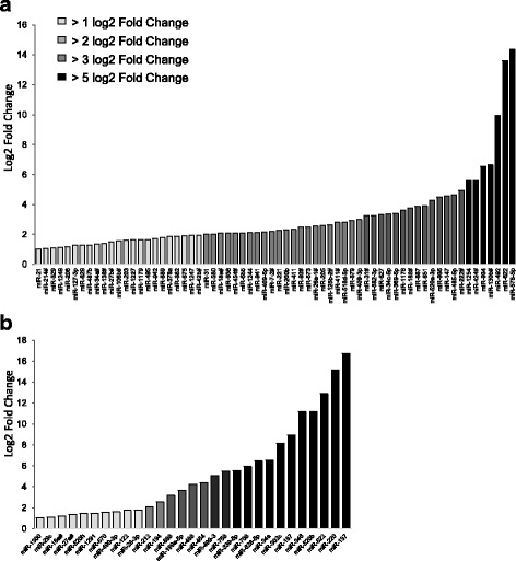
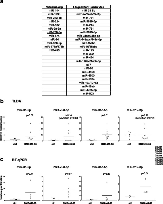
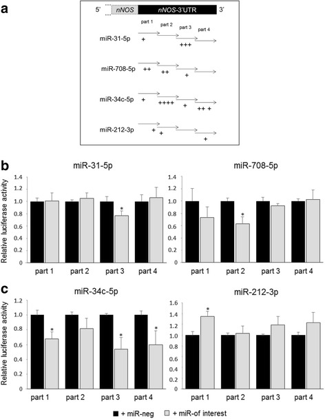
Methods: By a screening of 617 miRNAs in BMDd45-55 muscular biopsies using TLDA and an in silico study to determine which one could target nNOS, we selected four miRNAs. In order to select those that targeted a sequence of 3'UTR of NOS1, we performed luciferase gene reporter assay in HEK393T cells. Finally, expression of candidate miRNAs was modulated in control and DMD human myoblasts (DMDd45-52) to study their ability to target nNOS.

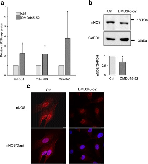
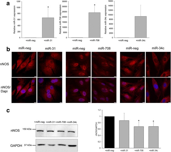
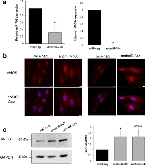
Results: TLDA assay and the in silico study allowed us to select four miRNAs overexpressed in muscle biopsies of BMDd45-55 compared to controls. Among them, only the overexpression of miR-31, miR-708, and miR-34c led to a decrease of luciferase activity in an NOS1-3'UTR-luciferase assay, confirming their interaction with the NOS1-3'UTR. The effect of these three miRNAs was investigated on control and DMDd45-52 myoblasts. First, we showed a decrease of nNOS expression when miR-708 or miR-34c were overexpressed in control myoblasts. We then confirmed that DMDd45-52 cells displayed an endogenous increased of miR-31, miR-708, and miR-34c and a decreased of nNOS expression, the same characteristics observed in BMDd45-55 biopsies. In DMDd45-52 cells, we demonstrated that the inhibition of miR-708 and miR-34c increased nNOS expression, confirming that both miRNAs can modulate nNOS expression in human myoblasts.

Conclusion: These results strongly suggest that miR-708 and miR-34c, overexpressed in dystrophic context, are new actors involved in the regulation of nNOS expression in dystrophic muscle.

Keywords: Becker muscular dystrophy (BMD); Duchenne muscular dystrophy (DMD); miRNA; nNOS.


Conflict of interest statement

Ethics approval and consent to participate

Muscle biopsies were collected from patients after informed consent, and this study was performed with agreement from the Committee for the Protection of Persons (CPP) concerned.

Competing interests

The authors declare that they have no competing interests.

Publisher’s Note

Springer Nature remains neutral with regard to jurisdictional claims in published maps and institutional affiliations.

Figures

Fig. 1
Fig. 1
Screening of miRNA expression profiling by TLDA in BMDd45-55 muscular biopsies. Data of TLDA were expressed by the value of Log2(R), where R is the ratio of the average of the relative quantification (RQ) obtained in BMDd45-55 muscles on the average of RQ values obtained from muscle of healthy subjects (a), or obtained from muscle of severe BMDd45-55 patients on the average of RQ values obtained from muscle of the moderate and mild BMDd45-55 patients (b), p ≤ 0.05. RQ are obtained using average of values of all miRNA for normalizer
Fig. 2
Fig. 2
Selection of candidate miRNAs. a In silico screening of miRNAs that could target NOS1 (TargetScan Human and microRNA.org). Candidate miRNAs are underlined. TLDA (Additional file 2, TLDA A2, B2) (b) and individual RT-qPCR (c) values of candidate miRNA expression in healthy subject biopsies (ctrl, black circle) and BMDd45-55 patients with asymptomatic phenotype (gray circle), moderate phenotype (gray square), severe phenotype (gray triangle), or not determined phenotype (gray hexagon); data are normalized on average of control expression. Lines represent average of each group. Individual RT-qPCR data are expressed as relative quantification using miR-30b as normalizer, normalized on average of control expression
Fig. 3
Fig. 3
miR-31, miR-708, and miR-34c targeted 3′UTR sequences of NOS1 gene. a Schematic positions of predicted binding sites by microT-CDS Diana Tools in 4 parts of 3′UTR of NOS1. b Relative luciferase activity of indicated miRNA-transfected cells normalized on luciferase activity in non-specific miRNA transfected cells (miR-neg). Cells were transfected with part 1, part 2, part 3, or part 4 of NOS1-3′UTR and with either miR-neg control (black bar) or miR of interest (gray bar). *p < 0.05
Fig. 4
Fig. 4
miR-31, miR-708, miR-34c, and nNOS expression in DMDd45-52 myoblasts. a miRNA expression in control human myoblasts and DMDd45-52 human myoblasts. Graph represents relative quantification of miRNA normalized on SNORD44 expression. miR-708 n = 7, miR-31 n = 7, and miR-34c n = 8. b nNOS immunoblot in control and DMDd45-52 cells. GAPDH serves as the loading control. Bar graph shows quantification results average of 8 independent experiments. c Control myoblasts immunolabeled with anti-nNOS (red) antibody, nuclei with Dapi (blue), and imaged by confocal microscopy. Scale bars, 10 μm. Representative of 3 independent experiments. *p ≤ 0.05
Fig. 5
Fig. 5
miR-708 and miR-34c overexpression inhibit nNOS expression in transfected control human myoblasts. a miRNA expression in control human myoblasts transfected with non-specific control miRNA (miR-neg, black bar) or indicated selected miRNA (gray bar). Graph represents average of relative quantification of miRNA normalized on SNORD44 expression of 5 (miR-31) or 3 (miR-708 and miR-34c) independent experiments. b Control myoblasts immunolabeled with anti-nNOS (red) antibody, nuclei with Dapi (blue), and imaged by confocal microscopy. Scale bars, 10 μm. Representative of 4 independent experiments. c nNOS immunoblot in transfected control human myoblasts. GAPDH serves as the loading control. Bar graph shows quantification results average of 5 independent experiments. *p ≤ 0.05
Fig. 6
Fig. 6
Inhibition of miR-708 and miR-34c increased nNOS expression in transfected DMDd45-52 human myoblasts. a miRNA expression in DMDd45-52 human myoblasts transfected with control non-specific miRNA (miR-neg, black bar) or indicated selected antimiR (gray bar). Graph represents average of relative quantification of miRNA normalized on SNORD44 expression of 6 (antimiR-708) or 3 (antimiR-34c) independent experiments. b DMDd45-52 myoblasts immunolabeled with anti-nNOS (red) antibody, nuclei with Dapi (blue), and imaged by confocal microscopy. Scale bars, 10 μm. Representative of 5 independent experiments. c nNOS immunoblot in transfected DMDd45-52 human myoblasts. GAPDH serves as the loading control. Bar graph shows quantification results average of 5 independent experiments. *p ≤ 0.05

References

    1. Monaco AP, Neve RL, Colletti-Feener C, Bertelson CJ, Kurnit DM, Kunkel LM. Isolation of candidate cDNAs for portions of the Duchenne muscular dystrophy gene. Nature. 1986;323:646–650. doi: 10.1038/323646a0. - DOI - PubMed
    1. Koenig M, Hoffman EP, Bertelson CJ, Monaco AP, Feener C, Kunkel LM. Complete cloning of the Duchenne muscular dystrophy (DMD) cDNA and preliminary genomic organization of the DMD gene in normal and affected individuals. Cell. 1987;50:509–517. doi: 10.1016/0092-8674(87)90504-6. - DOI - PubMed
    1. Monaco AP, Bertelson CJ, Liechti-Gallati S, Moser H, Kunkel LM. An explanation for the phenotypic differences between patients bearing partial deletions of the DMD locus. Genomics. 1988;2:90–95. doi: 10.1016/0888-7543(88)90113-9. - DOI - PubMed
    1. Koenig M, Beggs AH, Moyer M, Scherpf S, Heindrich K, Bettecken T, et al. The molecular basis for Duchenne versus Becker muscular dystrophy: correlation of severity with type of deletion. Am J Hum Genet. 1989;45:498–506. - PMC - PubMed
    1. Goyenvalle A, Davies KE. Engineering exon-skipping vectors expressing U7 snRNA constructs for Duchenne muscular dystrophy gene therapy. Methods Mol Biol. 2011;709:179–196. doi: 10.1007/978-1-61737-982-6_11. - DOI - PubMed

Publication types

MeSH terms

LinkOut - more resources