. 2018 Apr 27;15(1):79.
doi: 10.1186/s12985-018-0989-4.

Zika virus-induced hyper excitation precedes death of mouse primary neuron

Affiliations

Zika virus-induced hyper excitation precedes death of mouse primary neuron

Julie Gaburro et al. Virol J. .

Abstract

Background: Zika virus infection in new born is linked to congenital syndromes, especially microcephaly. Studies have shown that these neuropathies are the result of significant death of neuronal progenitor cells in the central nervous system of the embryo, targeted by the virus. Although cell death via apoptosis is well acknowledged, little is known about possible pathogenic cellular mechanisms triggering cell death in neurons.

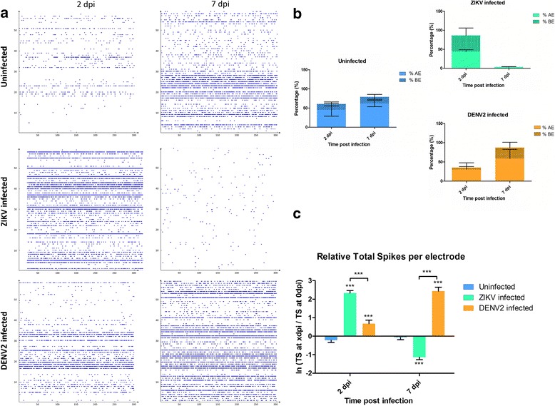
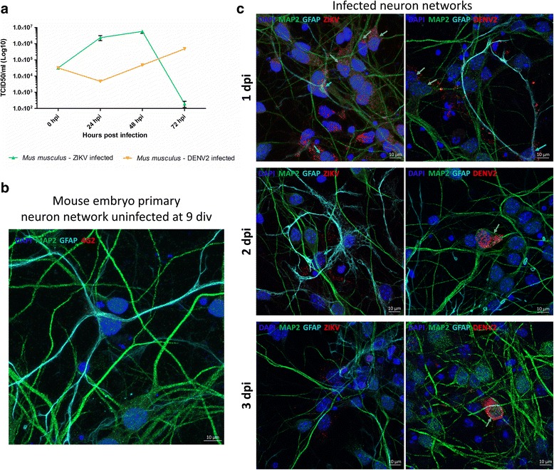
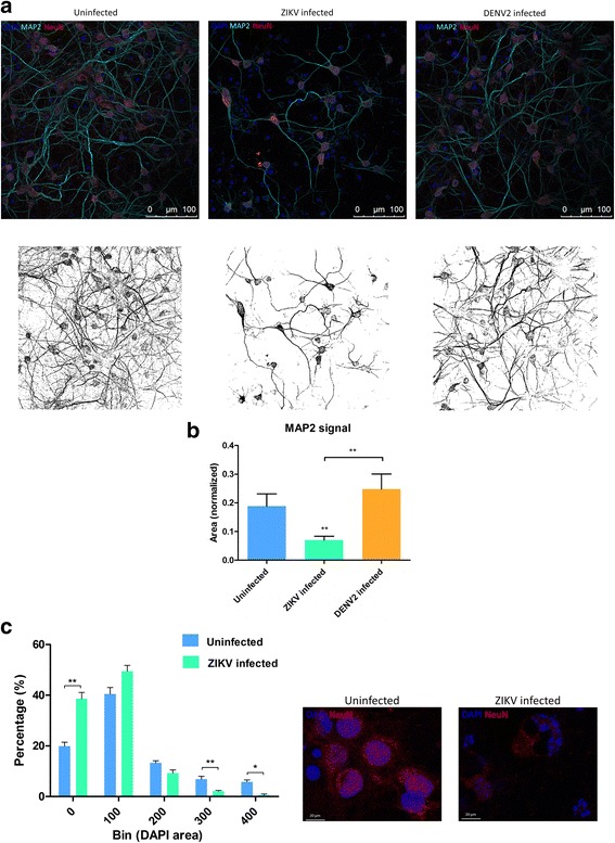
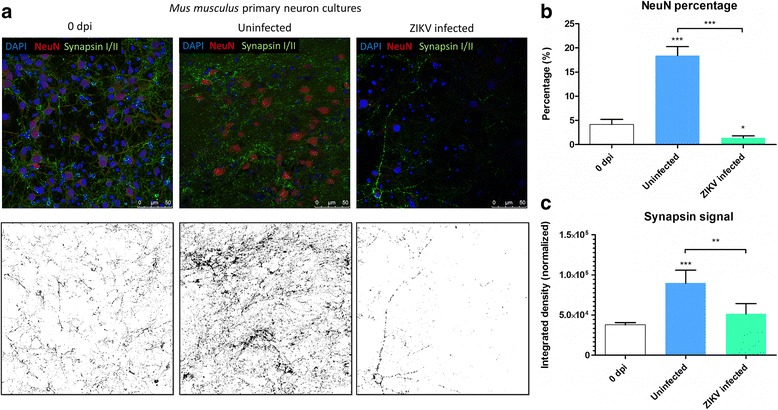
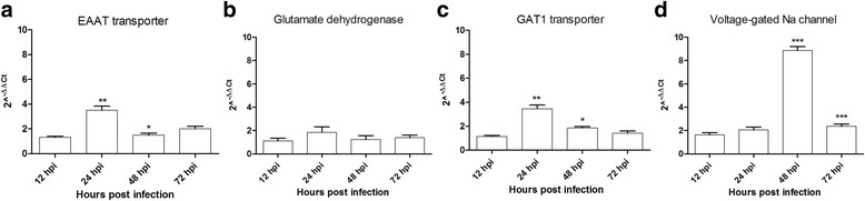
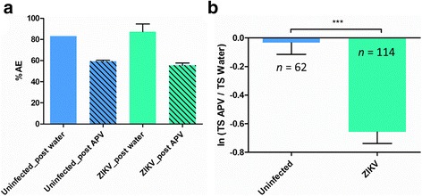
Methods: We used in vitro embryonic mouse primary neuron cultures to study possible upstream cellular mechanisms of cell death. Neuronal networks were grown on microelectrode array and electrical activity was recorded at different times post Zika virus infection. In addition to this method, we used confocal microscopy and Q-PCR techniques to observe morphological and molecular changes after infection.

Results: Zika virus infection of mouse primary neurons triggers an early spiking excitation of neuron cultures, followed by dramatic loss of this activity. Using NMDA receptor antagonist, we show that this excitotoxicity mechanism, likely via glutamate, could also contribute to the observed nervous system defects in human embryos and could open new perspective regarding the causes of adult neuropathies.

Conclusions: This model of excitotoxicity, in the context of neurotropic virus infection, highlights the significance of neuronal activity recording with microelectrode array and possibility of more than one lethal mechanism after Zika virus infection in the nervous system.

Keywords: Excitotoxicity; Glutamate; Hyper excitation; Microelectrode array; Primary neuron; Spike; Zika.


Conflict of interest statement

Competing interests

The authors declare that they have no competing interests.

Publisher’s Note

Springer Nature remains neutral with regard to jurisdictional claims in published maps and institutional affiliations.

Figures

Fig. 1
Fig. 1
Electrophysiological analysis of mouse embryo primary neurons spontaneous activity. a Raster plots of 5 min spiking activity of neuronal cultures on Microelectrode array (MEA) at different days post infection (dpi). b Percentages of active electrodes (AE) and bursting electrodes (BE) during spontaneous neuronal activity recording at different dpi of mouse primary neuron cultures. c Mouse spontaneous spiking activity with average ratio of total spike (TS) number per electrode between 0 dpi (reference) and TS number at 2 and 7 dpi of spontaneous activity. Statistical differences were calculated with unpaired t tests by comparing uninfected group to infected groups unless indicated by bracket. *P < 0.05; **P < 0.01; ***P < 0.001; means ± SEM. For c n values are detailed in Additional file 7: Table S3
Fig. 2
Fig. 2
Electrophysiological analysis of mouse embryo primary neurons under gabazine stimulus at 7dpi. a Mouse primary neurons stimulated activity with average ratio of TS number per electrode between gabazine stimulus and solvent (water) introduction; n = number of active electrodes. b Line charts representing the average relative change of the mean spike number per electrode between the reference (TC water) and the introduction of gabazine at different time points. Statistical differences were calculated with unpaired t tests by comparing uninfected group to infected groups unless indicated by bracket. *P < 0.05; **P < 0.01; ***P < 0.001; means ± SEM. c 3D electrode activity maps of the effect of gabazine stimulation mouse neuronal networks for 2 min, post water as baseline and post gabazine
Fig. 3
Fig. 3
Zika virus early infection dynamics at 1, 2 and 3 dpi. a Zika virus replication dynamics (using TCID50 in Vero cells) from 0 to 72 h post infection in mouse primary neurons (n = 3 per condition). b Confocal images of an uninfected mouse embryo neuron culture at 9 div (or 2 dpi). c Confocal images of ZIKV (left) or DENV2 (right) infected network at 1, 2 and 3 dpi, revealing the presence of virus (4G2 in red) in neurons (MAP2 in green) and glial cells (GFAP in turquoise)
Fig. 4
Fig. 4
Zika virus (ZIKV) effects on primary neuron networks from at 3 dpi. a Representative confocal images showing DAPI marker (blue), NeuN antibody (red), and MAP2 antibody (turquoise) at the top and its corresponding images with MAP2 staining below. Image analysis with ImageJ software reveals the differences of MAP2 signaling. b Quantification of MAP2 coverage, normalized by the number of DAPI counts in mouse primary neuron cultures at 3 dpi, n values are detailed in Additional file 2: Table S2. c Bar plots compare the size of DAPI stained nuclei of ZIKV infected and uninfected cells at 3 dpi. Each bin represents the distribution range size of nuclei in square pixel (n = 5 images for each condition). Bar plot values are shown with ± Standard Error of the Mean (SEM) with Mann-Whitney p-value on top, *P < 0.05; **P < 0.01; ***P < 0.001. On the right, confocal pictures illustrating nuclei sizes at 3 dpi
Fig. 5
Fig. 5
Zika virus (ZIKV) effects on primary neuron networks at 7 dpi. a Confocal images of mouse primary neuron culture before (0 dpi) and post ZIKV infection (7 dpi) at the top, respective Synapsin signal after applying image threshold with ImageJ at the bottom. b Percentage of mature mouse neurons stained with NeuN at 0 and 7 dpi, in mouse primary neuron cultures. c Quantification of Synapsin I/II signal, normalized by the number of DAPI counts on the right. Statistical differences were calculated with Mann-Whitney U tests, by comparing 0 dpi to the other groups unless indicated by bracket. *P < 0.05; **P < 0.01; ***P < 0.001; means ± SEM; n values are detailed in Additional file 2: Table S2
Fig. 6
Fig. 6
Genes mRNA expression after Zika virus infection in primary neuron cultures. Quantification of a Glutamate transporter (EEAT), b Glutamate dehydrogenase (GD1), c GABAA transporter (GAT1) and d Voltage-gated sodium channel (VGNaC) mRNA expression. Statistical differences were calculated with unpaired t-tests by comparing mRNA expression at timei to timei-1 within each group (n = 3). *P < 0.05; **P < 0.01; ***P < 0.001; means ± SEM
Fig. 7
Fig. 7
Microelectrode array (MEA) recording at 2 dpi after a NMDA-R antagonist introduction in mouse primary neuron cultures. a Percentage of Active Electrodes (AE) average from neuron network cultures after introduction of the NMDA-R antagonist APV (nMEA = 3 per condition). b Electrode activity calculated as average ratio of Total Spike (TS) number per electrode between APV and solvent introduction; Mann-Whitney U test, *P < 0.05; **P < 0.01; ***P < 0.001; means ± SEM; n = number of active electrodes

References

    1. Musso D, Nhan T, Robin E, Roche C, Bierlaire D, Zisou K, Shan Yan A, Cao-Lormeau V, Broult J. Potential for Zika virus transmission through blood transfusion demonstrated during an outbreak in French Polynesia, November 2013 to February 2014. Euro Surveill. 2014;19(14):20761. doi: 10.2807/1560-7917.ES2014.19.14.20761. - DOI - PubMed
    1. Oliveira Melo A, Malinger G, Ximenes R, Szejnfeld P, Alves Sampaio S, Bispo de Filippis A. Zika virus intrauterine infection causes fetal brain abnormality and microcephaly: tip of the iceberg? Ultrasound Obstet Gynecol. 2016;47(1):6–7. doi: 10.1002/uog.15831. - DOI - PubMed
    1. Mlakar J, Korva M, Tul N, Popović M, Poljšak-Prijatelj M, Mraz J, Kolenc M, Resman Rus K, Vesnaver Vipotnik T, Fabjan Vodušek V. Zika virus associated with microcephaly. N Engl J Med. 2016;2016(374):951–958. doi: 10.1056/NEJMoa1600651. - DOI - PubMed
    1. França GV, Schuler-Faccini L, Oliveira WK, Henriques CM, Carmo EH, Pedi VD, Nunes ML, Castro MC, Serruya S, Silveira MF. Congenital Zika virus syndrome in Brazil: a case series of the first 1501 livebirths with complete investigation. Lancet. 2016;388(10047):891–897. doi: 10.1016/S0140-6736(16)30902-3. - DOI - PubMed
    1. Cugola FR, Fernandes IR, Russo FB, Freitas BC, Dias JL, Guimarães KP, Benazzato C, Almeida N, Pignatari GC, Romero S. The Brazilian Zika virus strain causes birth defects in experimental models. Nature. 2016; - PMC - PubMed

MeSH terms

Substances