

Mutations in VP1 and 5'-UTR affect enterovirus 71 virulence
- PMID: 29703921
- PMCID: PMC5923339
- DOI: 10.1038/s41598-018-25091-7
Mutations in VP1 and 5'-UTR affect enterovirus 71 virulence
Erratum in
-
Author Correction: Mutations in VP1 and 5'-UTR affect enterovirus 71 virulence.Sci Rep. 2018 Jun 4;8(1):8744. doi: 10.1038/s41598-018-26833-3. Sci Rep. 2018. PMID: 29867107 Free PMC article.
Abstract
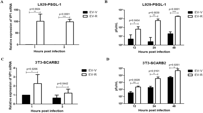
Enterovirus 71 (EV71) is a major cause of hand, foot and mouth disease (HFMD). The current EV71 propagating in Vero (EV-V) or sub-passaged in RD (EV-R) cells was used as a pathogen. Interestingly, EV-R exhibited differential virulence; challenging human scavenger receptor class B2-expressing (hSCARB2-Tg) mice with EV71 revealed that EV-V was more virulent than EV-R: 100% of mice that received lethal amounts of EV-V died, while all the mice that received EV-R survived. Severe pathogenesis correlated with viral burdens and proinflammatory cytokine levels were observed in EV-V-challenged mice, but controversy in EV-R-challenged mice. Consensus sequence analysis revealed EV-R rapidly acquired complete mutations at E145G and S241L and partial mutations at V146I of VP1, and acquired a T to C substitution at nucleotide 494 of the 5'-UTR. EV-R exhibited higher binding affinity for another EV71 receptor, human P-selectin glycoprotein ligand-1 (hPSGL-1), than EV-V. Both EV71s exhibited no significant difference in binding to hSCARB2. The molecular modelling indicate that these mutations might influence EV71 engagement with PSGL-1 and in vivo virulence.
Conflict of interest statement
The authors declare no competing interests.
Figures
Similar articles
-
Amino Acid Variation at VP1-145 of Enterovirus 71 Determines Attachment Receptor Usage and Neurovirulence in Human Scavenger Receptor B2 Transgenic Mice.J Virol. 2018 Jul 17;92(15):e00681-18. doi: 10.1128/JVI.00681-18. Print 2018 Aug 1. J Virol. 2018. PMID: 29848584 Free PMC article.
-
A Single Mutation in the VP1 Gene of Enterovirus 71 Enhances Viral Binding to Heparan Sulfate and Impairs Viral Pathogenicity in Mice.Viruses. 2020 Aug 13;12(8):883. doi: 10.3390/v12080883. Viruses. 2020. PMID: 32823486 Free PMC article.
-
A Single Mutation in the VP1 of Enterovirus 71 Is Responsible for Increased Virulence and Neurotropism in Adult Interferon-Deficient Mice.J Virol. 2016 Sep 12;90(19):8592-604. doi: 10.1128/JVI.01370-16. Print 2016 Oct 1. J Virol. 2016. PMID: 27440896 Free PMC article.
-
Recent Progress on Functional Genomics Research of Enterovirus 71.Virol Sin. 2019 Feb;34(1):9-21. doi: 10.1007/s12250-018-0071-9. Epub 2018 Dec 14. Virol Sin. 2019. PMID: 30552635 Free PMC article. Review.
-
Viral determinants that drive Enterovirus-A71 fitness and virulence.Emerg Microbes Infect. 2021 Dec;10(1):713-724. doi: 10.1080/22221751.2021.1906754. Emerg Microbes Infect. 2021. PMID: 33745413 Free PMC article. Review.
Cited by
-
Electrostatic interactions at the five-fold axis alter heparin-binding phenotype and drive enterovirus A71 virulence in mice.PLoS Pathog. 2019 Nov 15;15(11):e1007863. doi: 10.1371/journal.ppat.1007863. eCollection 2019 Nov. PLoS Pathog. 2019. PMID: 31730673 Free PMC article.
-
Heparan sulfate attachment receptor is a major selection factor for attenuated enterovirus 71 mutants during cell culture adaptation.PLoS Pathog. 2020 Mar 18;16(3):e1008428. doi: 10.1371/journal.ppat.1008428. eCollection 2020 Mar. PLoS Pathog. 2020. PMID: 32187235 Free PMC article.
-
Enterovirus A71 Vaccines.Vaccines (Basel). 2021 Feb 27;9(3):199. doi: 10.3390/vaccines9030199. Vaccines (Basel). 2021. PMID: 33673595 Free PMC article. Review.
-
SLC35B2 Acts in a Dual Role in the Host Sulfation Required for EV71 Infection.J Virol. 2022 May 11;96(9):e0204221. doi: 10.1128/jvi.02042-21. Epub 2022 Apr 14. J Virol. 2022. PMID: 35420441 Free PMC article.
-
Cellular receptors for enterovirus A71.J Biomed Sci. 2020 Jan 10;27(1):23. doi: 10.1186/s12929-020-0615-9. J Biomed Sci. 2020. PMID: 31924205 Free PMC article. Review.
References
Publication types
MeSH terms
Substances
LinkOut - more resources
Full Text Sources
Other Literature Sources
Molecular Biology Databases
Miscellaneous
