. 2019 Jan;56(1):378-393.
doi: 10.1007/s12035-018-1068-y. Epub 2018 Apr 27.

Targeted Gene Editing of Glia Maturation Factor in Microglia: a Novel Alzheimer's Disease Therapeutic Target

Affiliations

Targeted Gene Editing of Glia Maturation Factor in Microglia: a Novel Alzheimer's Disease Therapeutic Target

Sudhanshu P Raikwar et al. Mol Neurobiol. 2019 Jan.

Abstract

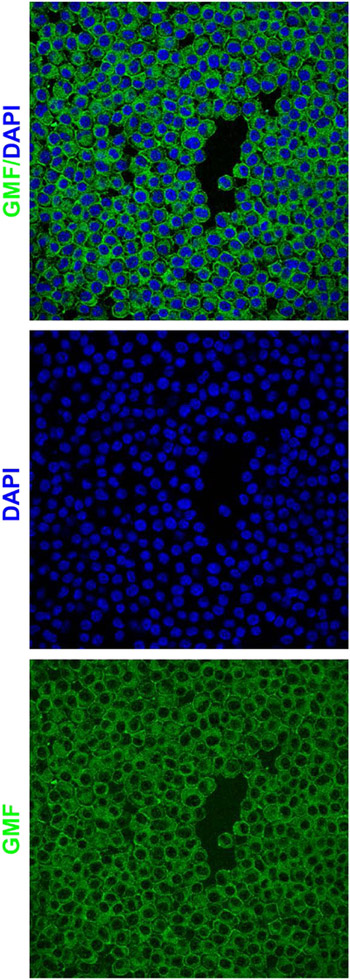
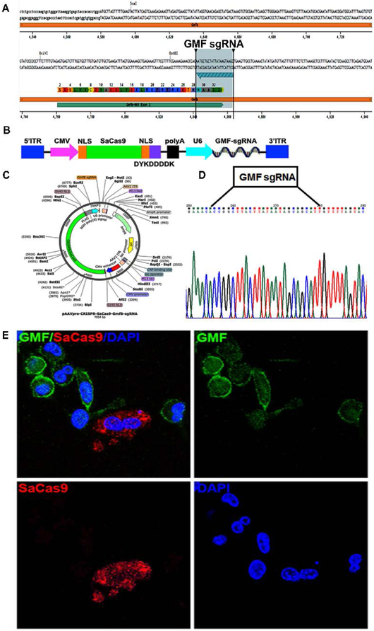
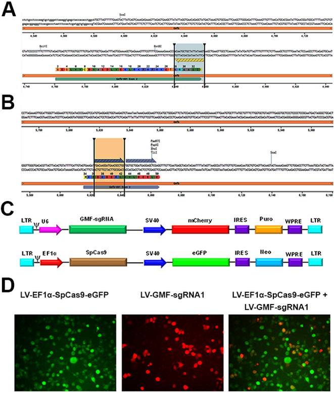
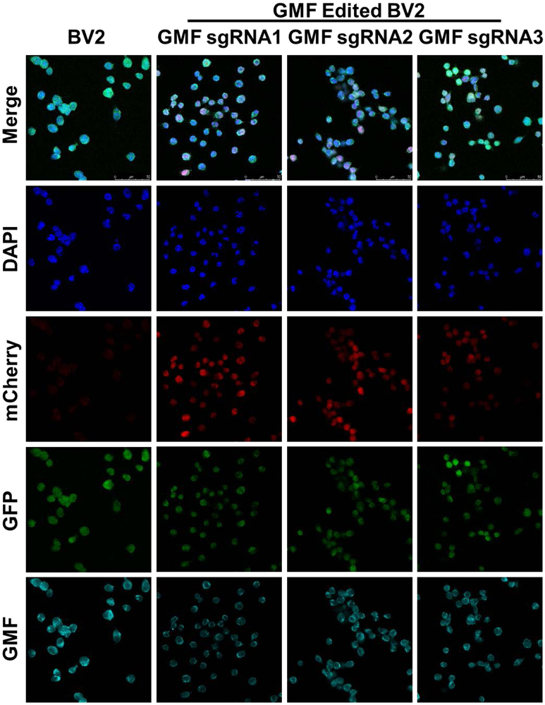
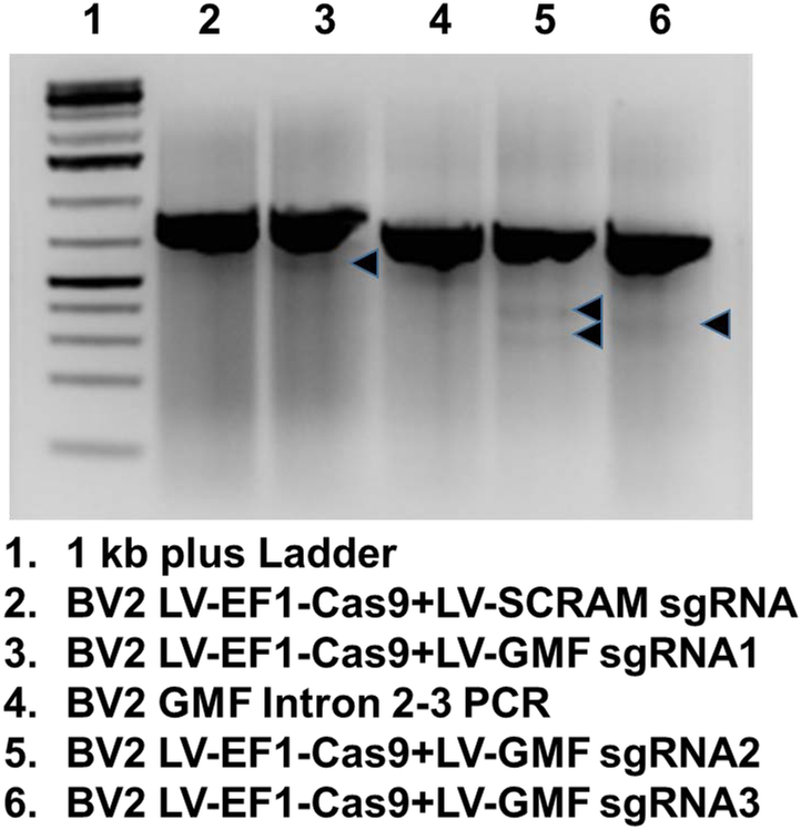
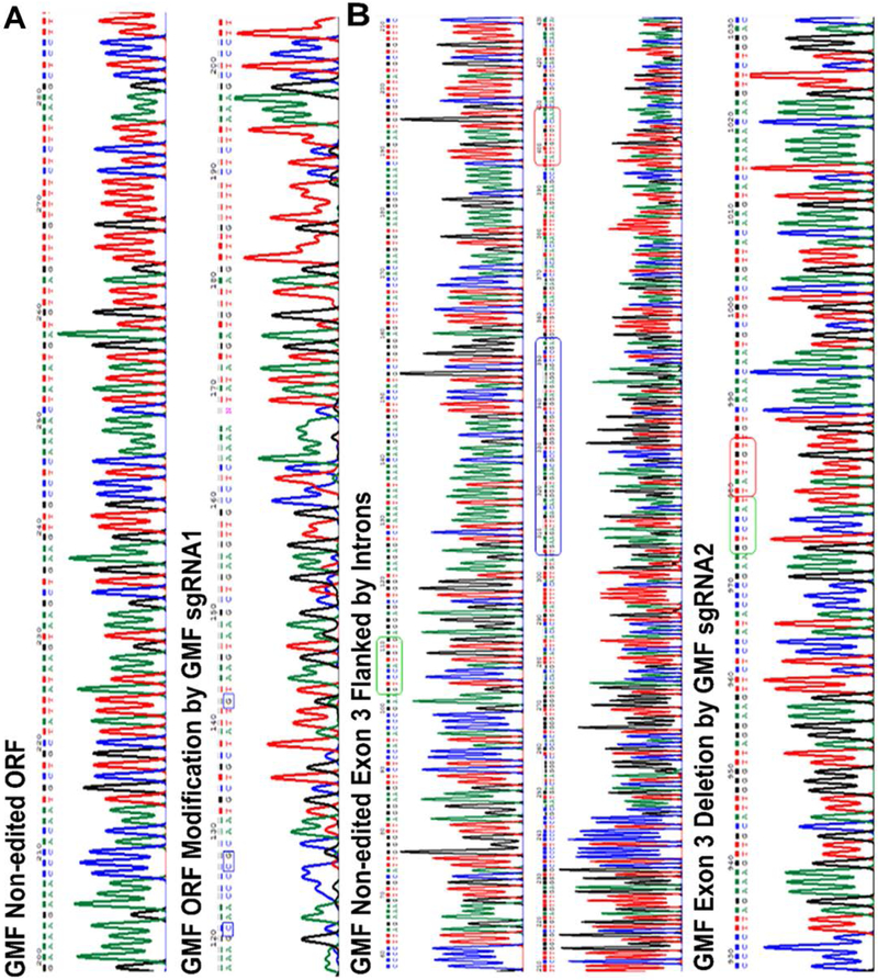
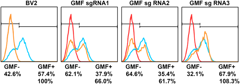
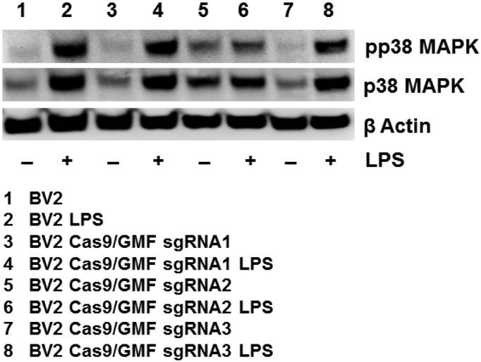
Alzheimer's disease (AD) is a devastating, progressive neurodegenerative disorder that leads to severe cognitive impairment in elderly patients. Chronic neuroinflammation plays an important role in the AD pathogenesis. Glia maturation factor (GMF), a proinflammatory molecule discovered in our laboratory, is significantly upregulated in various regions of AD brains. We have previously reported that GMF is predominantly expressed in the reactive glial cells surrounding the amyloid plaques (APs) in the mouse and human AD brain. Microglia are the major source of proinflammatory cytokines and chemokines including GMF. Recently clustered regularly interspaced short palindromic repeats (CRISPR) based genome editing has been recognized to study the functions of genes that are implicated in various diseases. Here, we investigated if CRISPR-Cas9-mediated GMF gene editing leads to inhibition of GMF expression and suppression of microglial activation. Confocal microscopy of murine BV2 microglial cell line transduced with an adeno-associated virus (AAV) coexpressing Staphylococcus aureus (Sa) Cas9 and a GMF-specific guide RNA (GMF-sgRNA) revealed few cells expressing SaCas9 while lacking GMF expression, thereby confirming successful GMF gene editing. To further improve GMF gene editing efficiency, we developed lentiviral vectors (LVs) expressing either Streptococcus pyogenes (Sp) Cas9 or GMF-sgRNAs. BV2 cells cotransduced with LVs expressing SpCas9 and GMF-sgRNAs revealed reduced GMF expression and the presence of indels in the exons 2 and 3 of the GMF coding sequence. Lipopolysaccharide (LPS) treatment of GMF-edited cells led to reduced microglial activation as shown by reduced p38 MAPK phosphorylation. We believe that targeted in vivo GMF gene editing has a significant potential for developing a unique and novel AD therapy.

Keywords: Adeno-associated virus; Alzheimer’s disease; CRISPR-Cas9; Glia maturation factor; Lentiviral vectors; Microglia.


Conflict of interest statement

Conflict of Interest

The authors confirm that they have no conflict of interest.

Figures

Figure 1:
Figure 1:. GMF is a novel AD therapeutic target for CRISPR-Cas9 based gene editing:
GMF immunostaining in the BV2 microglial cells indicates high-level perinuclear expression of GMF (Green) within the cytoplasm as well as on the cell surface. The nuclei (blue) are stained with DAPI. The images were acquired using the Leica TCP SP8 confocal microscope and processed using Leica Application Suite X software.
Figure 2:
Figure 2:. Development of AAV-SaCa9-GMF-sgRNA:
a) Selection of GMF sgRNA: The panel shows the DNA sequence of mouse GMF exon 2 flanked by the introns. The GMF amino acid sequence is indicated by single letter codes. The GMF sgRNA sequence located in the anticlockwise orientation on the lower strand is depicted by the green arrow flanked by the two vertical bars, b) Generation of AAV-SaCas9-GMF-sgRNA: The AAV ITRs flank the expression cassette comprising a CMV promoter driving the expression of a nuclear localized SaCas9 which also harbors the DYKDDDK tag, followed by a polyA signal. The second expression casette bears a U6 promoter driven GMF sgRNA, c) Detailed vector map depicting various elements present in pAAVpro-CRISPR-SaCas9-GMF-sgRNA, d) The DNA sequence of the GMF sgRNA cloned into the AAV vector is depicted flanked by the sequence of the expression cassette, e) Targeted GMF gene editing mediated by AAV-SaCas9-GMF-sgRNA in BV2 cells. The immunostaining for the GMF (green) and the DYKDDDK tag (red) indicates that the non transduced BV2 cells display significant intracellular GMF expression with higher concentrations towards the cell periphery and on the plasma membrane. The GMF non-edited cells do not display any red color and are therefore negative for SaCas9 expression. GMF gene edited cells do not exhibit GMF expression and are higly positive for SaCas9 expression thereby indicating biallelic GMF gene editing mediated by SaCas9 and GMF-sgRNA.
Figure 3:
Figure 3:. Development of LV-EF1α-SpCas9-eGFP and LV-GMF-sgRNAs:
a) Selection of GMF specific sgRNAs: The panel A shows the DNA sequence of mouse GMF exon 2 flanked by the introns. The GMF amino acid sequence is indicated by single letter codes. The GMF sgRNA1 sequence which is slightly different than the GMF sgRNA used in the AAV (due to the differences between SaCas9 and SpCas9 PAM sequences) is also located in the anticlockwise orientation on the lower strand is depicted by the blue arrow flanked by the two vertical bars. b) The panel B shows the GMF sgRNA deisgn of GMF sgRNA2 and GMF sgRNA3 both of which are located in the GMF coding sequence in the exon 3. c) Generation of LV-EF1α-SpCas9-eGFP-Neo and LV-GMF-sgRNAs: A set of 5 different VSV enveloped pseudotyped third generation lentiviral vectors were developed. In the lentiviral vector LV-SpCas9-eGFP-Neo, the EF1α promoter drives the expression of CRISPR-Cas9 and a SV40 promoter drives the expression of eGFP reporter and Neo resistance genes. The lentiviral vector LV-GMF-sgRNA 1, 2 and 3 express GMF sgRNAs under the control of the U6 promoter while the SV40 promoter drives the expression of mCherry reporter and Puromycin resistance marker. In the control lentiviral vector LV-SCRAM-sgRNA a scrambled sgRNA that does not target any known gene was used as a negative gene editing control virus. d) Generation of stable CRISPR-Cas9 expressing cell line: BV2 cells were transduced with the lentiviral vector LV-EF1α-SpCas9-eGFP-Neo to generate a stable cell line using neomycin selection. Co-transduction of BV2-CRISPR-Cas9 cells with LV-GMF-sgRNA 1 shows the presence of mCherry expressing cells and the merged image shows the presence of a small subset of cells showing both eGFP and mCherry expression. Puromycin selection was used to generate stable BV2-CRISPR-Cas9 and GMF-sgRNA expressing cells.
Figure 4:
Figure 4:. Targeted GMF gene editing using LV-EF1α-SpCas9-eGFP+LV-GMF-sgRNAs:
a) Confocal microscopy of the BV2-CRISPR-Cas9 cells expressing SpCas9 (Green) reveal normal GMF expression (Cyan) with DAPI stained nuclei (Blue) in the far left vertical panel labeled BV2. b) Co-transduction of BV2-CRISPR-Cas9 cells with GMF-specific sgRNA1 leads to a differential GMF editing and partial reduction in GMF expression in the vertical panel labeled as GMF sgRNA1. c) Co-transduction of BV2-CRISPR-Cas9 cells with GMF-specific sgRNA2 leads to a differential GMF editing and partial reduction in GMF expression in the vertical panel labeled GMF sgRNA2. d) Co-transduction of BV2-CRISPR-Cas9 cells with GMF-specific sgRNA3 leads to a differential GMF editing and partial reduction in GMF expression in the vertical panel labeled as GMF sgRNA3. In the panels depicting GMF sgRNAs 1-3, mCherry expression indicates co-expression of GMF sgRNAs. The GMF gene edited BV2 cells with reduced GMF expression are marked with white filled triangles.
Figure 5:
Figure 5:. Mutational Analysis reveals GMF gene editing in BV2 cells:
The genomic DNA from the non edited or GMF edited cells was isolated and used for PCR amplification of the GMF edited sequence. The PCR amplified products from the wild type BV2 cells as well as from the GMF edited cells were used for heteroduplex formation and subjected to treatment with Guide-it Resolvase and subsequently analyzed for the cleavage of the mismatched DNA. The DNA mixture was resolved by agarose gel electrophoresis. The DNA in the lanes 2 and 4 does not reveal any cleavage as expected. However, the DNA in the lanes 3, 5 and 6 indicate low levels of DNA cleavage indicating successful GMF gene editing. The faint bands resulting due to Guide-it Resolvase-mediated cleavage are shown as black arrows.
Figure 6A:
Figure 6A:. SpCas9-mediated GMF editing leads to indels in GMF coding sequence:
a) The top panel shows the wild type GMF DNA sequence while the bottom panel shows the GMF edited sequence (using GMF-sgRNA1) from the exon 2 and is flanked by the intron sequences. GMF edited DNA sequence as a result of GMF sgRNA1-mediated editing leads to indels in the GMF coding sequence causing a frameshift in the GMF coding sequence. Specific nucleotide changes are indicated in the blue box, b) The top and the middle panels show the normal non-edited DNA sequence of the GMF exon 3 and the flanking introns. The bottom panel shows the deletion of GMF exon 3 post GMF gene editing. There is a significant change in the nucleotide sequence as a result of GMF gene editing. The green and the red boxes in the top and the middle panels depict the normal GMF DNA sequence. The GMF exon 3 is depicted within the blue box in the middle panel. As a result of GMF gene editing the GMF exon 3 and the flanking partial intronic sequences have been deleted and are represented as green and red boxes in the bottom panel.
Figure 7:
Figure 7:. Despite stable expression of SpCas9 and GMF sgRNAs, GMF editing is moderate:
Flow cytometric analysis of GMF gene editing in BV2 cells revealed that as compared to the normal GMF expression in BV2 cells, GMF gene editing leads to significant reduction in GMF expression especially due to GMF sgRNA1 and sgRNA2. Surprisingly there was no significant change in GMF expression due to GMF sgRNA3-mediated GMF gene editing. The changes in GMF expression are indicated in terms of GMF positive and GMF negative cell populations.
Figure 8:
Figure 8:. GMF gene editing causes suppression of MAPK pathway activation:
Wild type BV2 cells, and GMF gene edited BV2 cells were treated with 1.0μg/ml LPS for 30 minutes and analyzed for the expression of pp38MAPK and p38 MAPK. The representative western blot data (n=3) indicates that wild type BV2 cells display enhanced pp38 MAPK and p38 MAPK expression in the presence of LPS treatment (lanes 1 and 2). However, GMF sgRNA2-edited BV2 cells exhibit significant reduction in both pp38 MAPK and p38 MAPK expression with and without LPS treatment (lanes 5 and 6). Surprisingly, pp38 MAPK and p38 MAPK expression patterns are very similar between wild type as well as GMF sgRNA1 and sgRNA3-edited BV2 cells as depicted in lanes 3, 4 and 7, 8 respectively.

References

    1. Doody RS, Farlow M, Aisen PS, Alzheimer's Disease Cooperative Study Data A, Publication C (2014) Phase 3 trials of solanezumab and bapineuzumab for Alzheimer's disease. N Engl J Med 370 (15):1460. doi: 10.1056/NEJMc1402193 - DOI - PubMed
    1. Doody RS, Thomas RG, Farlow M, Iwatsubo T, Vellas B, Joffe S, Kieburtz K, Raman R, Sun X, Aisen PS, Siemers E, Liu-Seifert H, Mohs R, Alzheimer's Disease Cooperative Study Steering C, Solanezumab Study G (2014) Phase 3 trials of solanezumab for mild-to-moderate Alzheimer's disease. N Engl J Med 370 (4):311–321. doi: 10.1056/NEJMoa1312889 - DOI - PubMed
    1. Honig LS, Vellas B, Woodward M, Boada M, Bullock R, Borrie M, Hager K, Andreasen N, Scarpini E, Liu-Seifert H, Case M, Dean RA, Hake A, Sundell K, Poole Hoffmann V, Carlson C, Khanna R, Mintun M, DeMattos R, Selzler KJ, Siemers E (2018) Trial of Solanezumab for Mild Dementia Due to Alzheimer's Disease. N Engl J Med 378 (4):321–330. doi: 10.1056/NEJMoa1705971 - DOI - PubMed
    1. Sevigny J, Chiao P, Bussiere T, Weinreb PH, Williams L, Maier M, Dunstan R, Salloway S, Chen T, Ling Y, O'Gorman J, Qian F, Arastu M, Li M, Chollate S, Brennan MS, Quintero-Monzon O, Scannevin RH, Arnold HM, Engber T, Rhodes K, Ferrero J, Hang Y, Mikulskis A, Grimm J, Hock C, Nitsch RM, Sandrock A (2016) The antibody aducanumab reduces Abeta plaques in Alzheimer's disease. Nature 537 (7618):50–56. doi: 10.1038/nature19323 - DOI - PubMed
    1. Graham WV, Bonito-Oliva A, Sakmar TP (2017) Update on Alzheimer's Disease Therapy and Prevention Strategies. Annu Rev Med 68:413–430. doi: 10.1146/annurev-med-042915-103753 - DOI - PubMed

Substances