Review
. 2019 Jan;97(1):45-56.
doi: 10.1002/jnr.24250. Epub 2018 Apr 28.

Insights into optic pathway glioma vision loss from mouse models of neurofibromatosis type 1

Affiliations
Review

Insights into optic pathway glioma vision loss from mouse models of neurofibromatosis type 1

Morgan E Freret et al. J Neurosci Res. 2019 Jan.

Abstract

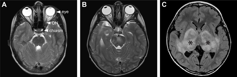
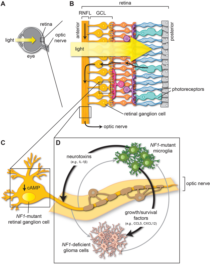
Neurofibromatosis type 1 (NF1) is a common cancer predisposition syndrome caused by mutations in the NF1 gene. The NF1-encoded protein (neurofibromin) is an inhibitor of the oncoprotein RAS and controls cell growth and survival. Individuals with NF1 are prone to developing low-grade tumors of the optic nerves, chiasm, tracts, and radiations, termed optic pathway gliomas (OPGs), which can cause vision loss. A paucity of surgical tumor specimens and of patient-derived xenografts for investigative studies has limited our understanding of human NF1-associated OPG (NF1-OPG). However, mice genetically engineered to harbor Nf1 gene mutations develop optic gliomas that share many features of their human counterparts. These genetically engineered mouse (GEM) strains have provided important insights into the cellular and molecular determinants that underlie mouse Nf1 optic glioma development, maintenance, and associated vision loss, with relevance by extension to human NF1-OPG disease. Herein, we review our current understanding of NF1-OPG pathobiology and describe the mechanisms responsible for tumor initiation, growth, and associated vision loss in Nf1 GEM models. We also discuss how Nf1 GEM and other preclinical models can be deployed to identify and evaluate molecularly targeted therapies for OPG, particularly as they pertain to future strategies aimed at preventing or improving tumor-associated vision loss in children with NF1.

Keywords: RAS; brain tumor; neurofibromatosis type 1; neurofibromin; optic nerve; optic pathway glioma; retinal ganglion cell; vision loss.


Conflict of interest statement

Conflict of interest: The authors declare no conflicts of interest

Figures

Figure 1.
Figure 1.. Location and MRI characteristics of NF1-optic pathway gliomas (OPGs).
(A) Axial T2-weighted MRI scan depicts a normal optic chiasm and optic nerves (ONs) in a child with NF1 lacking an OPG. (B) Axial T2-weighted MRI scan shows an OPG in a child with NF1 involving the optic chiasm and nerves. The chiasm is enlarged and diffusely hyperintense. The optic nerves show fusiform enlargement and tortuosity bilaterally (arrowheads). (C) Axial T1-weighted non-contrast MRI scan in a different child with NF1 with an OPG involving the optic radiations (asterisks).
Figure 2.
Figure 2.. RAS effector pathway de-regulation underlies optic pathway glioma (OPG) growth and tumor-associated retinal ganglion cell (RGC) dysfunction.
Neurofibromin is a negative regulator of the RAS proto-oncogene product. It accelerates the conversion of active RAS-GTP to inactive GDP-bound RAS. (A) In tumor cells with biallelic NF1 inactivation (NF1-deficient cells), there is increased activation of the MEK-ERK and PI3K-AKT-mTOR pathways, resulting in greater cell proliferation and survival. RAS hyperactivation also leads to decreased cyclic AMP (cAMP) levels to promote tumor cell survival. (B) In RGCs heterozygous for a germline NF1 gene mutation (NF1-mutant cells), impaired neurofibromin function leads to reduced adenylyl cyclase-mediated cAMP production and increased RGC death. Based on mechanistic studies in other CNS neurons, the reduced cAMP in RGCs likely results from increased RAS-dependent activation of protein kinase C-ζ (PKCζ).
Figure 3.
Figure 3.. Cell-autonomous and cell-non-autonomous mechanisms underlying optic pathway glioma pathogenesis and associated vision loss.
(A, B) Sagittal sections through the mammalian eye (A) and retina (B). Photoreceptor (cone and rod) afferent pathways converge on retinal ganglion cells (RGCs). RGC bodies reside in the ganglion cell layer (GCL) of the retina, and their axons course through the retinal nerve fiber layer (RNFL) into the optic nerve. (C) In NF1-mutant RGCs, impaired neurofibromin function reduces intracellular cAMP levels, thereby lowering the threshold for RGC death and subsequent vision loss. (D) NF1-mutant microglia secrete chemokines (e.g., CCL5, CXCL12), which promote the proliferation and survival of NF1-deficient tumor cells. In addition, estrogen receptor β (ERβ)-mediated microglial priming leads to the production of neurotoxins (e.g., IL-1β) that increase NF1-mutant RGC axonal dysfunction and death, causing vision loss in a sex-dependent manner. Tan-colored cells denote NF1-mutant oligodendrocytes that ensheath the optic nerve axons. The gray arrows indicate potential intercellular interactions in NF1-OPG, which are described in the main text.

References

    1. Alvord EC Jr., Lofton S. 1988. Gliomas of the optic nerve or chiasm. Outcome by patients’ age, tumor site, and treatment. J Neurosurg 68(1):85–98. - PubMed
    1. Anastasaki C, Gutmann DH. 2014. Neuronal NF1/RAS regulation of cyclic AMP requires atypical PKC activation. Hum Mol Genet 23(25):6712–6721. - PMC - PubMed
    1. Anastasaki C, Le LQ, Kesterson RA, Gutmann DH. 2017. Updated nomenclature for human and mouse neurofibromatosis type 1 genes. Neurol Genet 3(4):e169. - PMC - PubMed
    1. Anastasaki C, Woo AS, Messiaen LM, Gutmann DH. 2015. Elucidating the impact of neurofibromatosis-1 germline mutations on neurofibromin function and dopamine-based learning. Hum Mol Genet 24(12):3518–3528. - PMC - PubMed
    1. Andreou KE, Soto MS, Allen D, Economopoulos V, de Bernardi A, Larkin JR, Sibson NR. 2017. Anti-inflammatory Microglia/Macrophages As a Potential Therapeutic Target in Brain Metastasis. Front Oncol 7:251. - PMC - PubMed

Publication types