Review
. 2018 Jul:90:247-259.
doi: 10.1016/j.neubiorev.2018.04.014. Epub 2018 Apr 25.

The neuroanatomic complexity of the CRF and DA systems and their interface: What we still don't know

Affiliations
Review

The neuroanatomic complexity of the CRF and DA systems and their interface: What we still don't know

E A Kelly et al. Neurosci Biobehav Rev. 2018 Jul.

Abstract

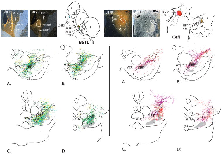
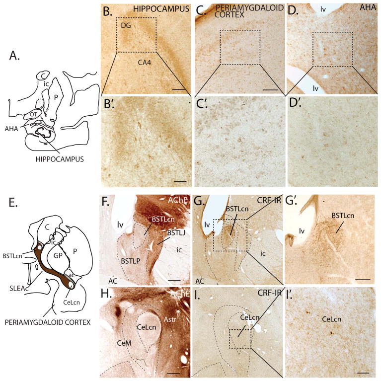
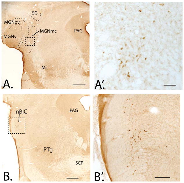
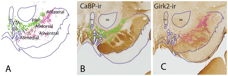
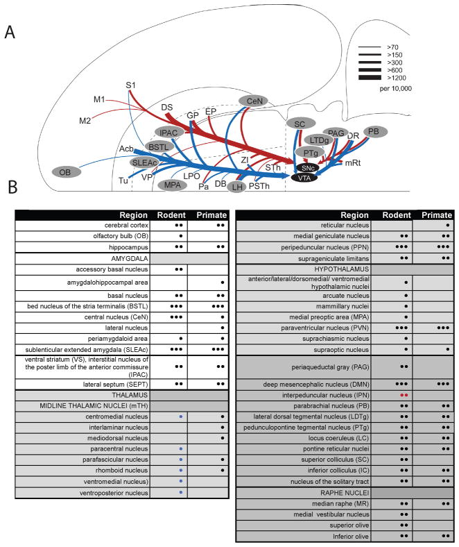
Corticotropin-releasing factor (CRF) is a neuropeptide that mediates the stress response. Long known to contribute to regulation of the adrenal stress response initiated in the hypothalamic-pituitary axis (HPA), a complex pattern of extrahypothalamic CRF expression is also described in rodents and primates. Cross-talk between the CRF and midbrain dopamine (DA) systems links the stress response to DA regulation. Classically CRF + cells in the extended amygdala and paraventricular nucleus (PVN) are considered the main source of this input, principally targeting the ventral tegmental area (VTA). However, the anatomic complexity of both the DA and CRF system has been increasingly elaborated in the last decade. The DA neurons are now recognized as having diverse molecular, connectional and physiologic properties, predicted by their anatomic location. At the same time, the broad distribution of CRF cells in the brain has been increasingly delineated using different species and techniques. Here, we review updated information on both CRF localization and newer conceptualizations of the DA system to reconsider the CRF-DA interface.

Keywords: Extended amygdala; Mouse; Primate; Rat; Retrorubral field; Stress; Substantia nigra; VTA.


Figures

Figure 1
Figure 1
Anterogradely labeled fibers resulting from all injections into the primate BSTL (A–D) and CeN (A′–D′) terminate mainly over the parabrachial pigmented nucleus (PBP), the lateral-most wing of the A10 neurons, and A8 (RRF), with some encroachment on the A9 neurons (gray). The VTA is receives fewer inputs. Photomicrographs show representative tracer injection sites the BSTL (J28LY) and CeN (J1FR) with adjacent sections labeled for either somatostatin (J28SST) or TH (J1TH) to localize relative injection site placement with nuclear subdivisions. Abbreviations: FR, fluororuby conjugated dextran amine; FS, fluorescein conjugated to dextran amine; LY, Lucifer yellow conjugated to dextran amine. Scale bar, 500 μm. *Figure reproduced with permission from (Fudge et al 2017)
Figure 2
Figure 2
A–D, B′–D′: CRF-IR cells in regions of monkey hippocampus and surrounding amygdala transition areas. B–B′. CRF-IR cells CA4 field of the hippocampus at low (B) and higher power (B′). C–C′. CRF-IR cells in the periamygdaloid cortex at low (C) and high (C′) power, and amygdalohippocampal area (AHA) at low (D) and high power (D′). E. Schematic showing the central extended amygdala. F. Subdivisions of the BSTL based on AChE staining. G–G′: CRF-IR cells and fibers stand out in the BSTLcn both at low (G) and high (G′) power. H. Subdivisions of the CeN in AChE. I–I′: Relatively low concentrations of CRF-IR cells and fibers in the CeLcn at low (I) and high magnification (I′) compared to the BSTLcn (G–G′). Abbreviations: AC; anterior commissure; AChE, acetylcholinesterase; AHA, amygdalohippocampal area; Astr, amygdalostriatal area; BSTLcn, bed nucleus of the stria terminalis, lateral central division; BSTLJ, bed nucleus of the stria terminalis, lateral juxtacapsular division; BSTLP, bed nucleus of the stria terminalis, lateral posterior division; C, caudate nucleus; CeLcn, central nucleus, lateral central division; CeM, central nucleus, medial subdivision; DG; dentate gyrus; GP, globus pallidus; ic, internal capsule; lv, lateral ventricle; OT, optic tract; P, putamen; PAC, periamygdaloid cortex; SLEAc, sublenticular extended amygdala, central subdivision. Scale bars: = C, F, G, H, I = 500 μm; G′=250 μm; B, D and I′ = 100 μm; 40X magnification in B′, C′, D′ =50 μm.
Figure 3
Figure 3
A. Low power photomicrograph of CRF-IR cells in monkey medial geniculate nucleus, magnocellular division (MGNmc), extending dorsally into the suprageniculate nucleus (SG). Subdivisions determined using adjacent sections immunostained for calbindin-D28k, not shown. A′. Higher power image of CRF-labeled cells in the MGNmc (boxed area of the in A). B. At slightly caudal levels of the pons, CRF-IR cells in the brachial nucleus of the inferior colliculus (nBIC), with higher magnification in B′. Abbreviations: ICnb; brachial nucleus of the inferior colliculus; ML, medial lemniscus; MGNpv, medial geniculate nucleus, posteroventral subdivision; MGNv, medial geniculate nucleus, ventral subdivision; PAG, periaqueductal gray; PTg, pedunculopontine tegmental nucleus; SCP, superior cerebellar peduncle. Scale bars: A and B= 1mm; A′= 100 μm, B′= 250 μm.
Figure 4
Figure 4
A. Subdivisions of the primate ventral midbrain dopamine (DA) neurons depicted in adjacent sections. B. CalbindinD-28k (CaBP) positive cells populate the VTA and PBP (green). Note that the A9 cells are devoid of CaBP-IR. C. G protein-coupled inwardly-rectifying potassium channel 2 (Girk2) labeled cells (pink) are largely aggregated in the A9 neurons, which are further divided into medial, lateral, dorsal, and ventral subregions. Charts of CaBP-IR and Girk2-IR cells are overlaid in A, matching landmarks. Abbreviations: IIIn, third nerve; RN, red nucleus.
Figure 5
Figure 5
A–B. Dark-field photomicrographs showing CRF-IR fibers in the caudal midbrain. A. Labeled fibers are densest in the retrorubral field (RRF, or A8) with fewer labeled fibers in the ventral tegmental area (VTA, A10). B. Higher power image of RRF (boxed area in A). IPN, interpeduncular nucleus. Scale bar= 250 μm.
Figure 6
Figure 6
A. Many of the known afferent sources to the midbrain dopamine neurons, adapted from (Watabe-Uchida et al 2012) with permission. Blue indicates predominant inputs to VTA while red indicates predominant inputs to A9 dopamine neurons, in mouse. gray-highlighted regions depict brain regions that also contain CRF-IR cells. Dopamine afferent sources from DS to VTA and Acb to SNc were omitted for clarity. Line thickness corresponds to the number of input neurons in each area (inputs per 10,000 total inputs; per Watabe-Uchida et al 2012). B. Relative distribution of CRF cells in rodent and primate. • = few, •• = moderate, ••• = robust as translated from the literature. For rodent, blue symbols correspond to “rat only”, red symbols correspond to “mouse only”. Abbreviations not shown in the Table: DB, diagonal band of Broca; DS, dorsal striatum; EP, endopeduncular nucleus; GP, globus pallidus; LPO, lateral preoptic area; M1, M2, primary and secondary motor cortex; PSTh, parasubthalamic nuclei; S1, primary sensory cortex; VP, ventral pallidum; ZI, zona incerta; sTH, subthalamic nuclei.

References

    1. Adamantidis AR, Tsai HC, Boutrel B, Zhang F, Stuber GD, et al. Optogenetic interrogation of dopaminergic modulation of the multiple phases of reward-seeking behavior. J Neurosci. 2011;31:10829–35. - PMC - PubMed
    1. Alon T, Zhou L, Perez CA, Garfield AS, Friedman JM, Heisler LK. Transgenic mice expressing green fluorescent protein under the control of the corticotropin-releasing hormone promoter. Endocrinology. 2009;150:5626–32. - PMC - PubMed
    1. Anderegg A, Poulin JF, Awatramani R. Molecular heterogeneity of midbrain dopaminergic neurons--Moving toward single cell resolution. FEBS Lett. 2015;589:3714–26. - PMC - PubMed
    1. Antoni FA, Palkovits M, Makara GB, Linton EA, Lowry PJ, Kiss JZ. Immunoreactive corticotropin-releasing hormone in the hypothalamoinfundibular tract. Neuroendocrinology. 1983;36:415–23. - PubMed
    1. Asan E, Yilmazer-Hanke DM, Eliava M, Hantsch M, Lesch KP, Schmitt A. The corticotropin-releasing factor (CRF)-system and monoaminergic afferents in the central amygdala: investigations in different mouse strains and comparison with the rat. Neuroscience. 2005;131:953–67. - PubMed

Publication types

MeSH terms