. 2018 Jul;142(1):235-245.e6.
doi: 10.1016/j.jaci.2018.02.053. Epub 2018 Apr 27.

Transfer of gene-corrected T cells corrects humoral and cytotoxic defects in patients with X-linked lymphoproliferative disease

Affiliations

Transfer of gene-corrected T cells corrects humoral and cytotoxic defects in patients with X-linked lymphoproliferative disease

Neelam Panchal et al. J Allergy Clin Immunol. 2018 Jul.

Abstract

Background: X-linked lymphoproliferative disease 1 arises from mutations in the SH2D1A gene encoding SLAM-associated protein (SAP), an adaptor protein expressed in T, natural killer (NK), and NKT cells. Defects lead to abnormalities of T-cell and NK cell cytotoxicity and T cell-dependent humoral function. Clinical manifestations include hemophagocytic lymphohistiocytosis, lymphoma, and dysgammaglobulinemia. Curative treatment is limited to hematopoietic stem cell transplantation, with outcomes reliant on a good donor match.

Objectives: Because most symptoms arise from defective T-cell function, we investigated whether transfer of SAP gene-corrected T cells could reconstitute known effector cell defects.

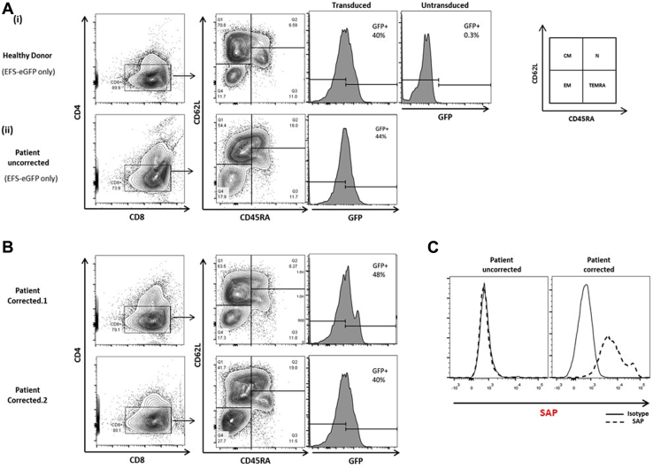
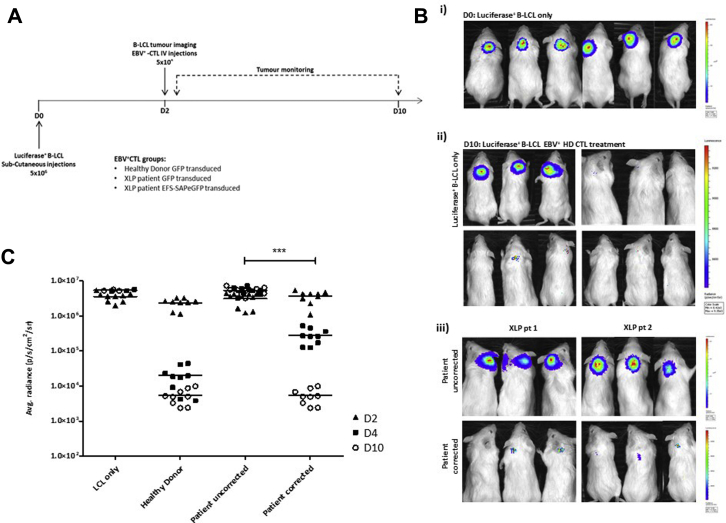
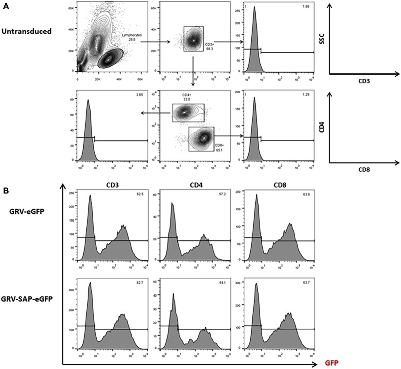
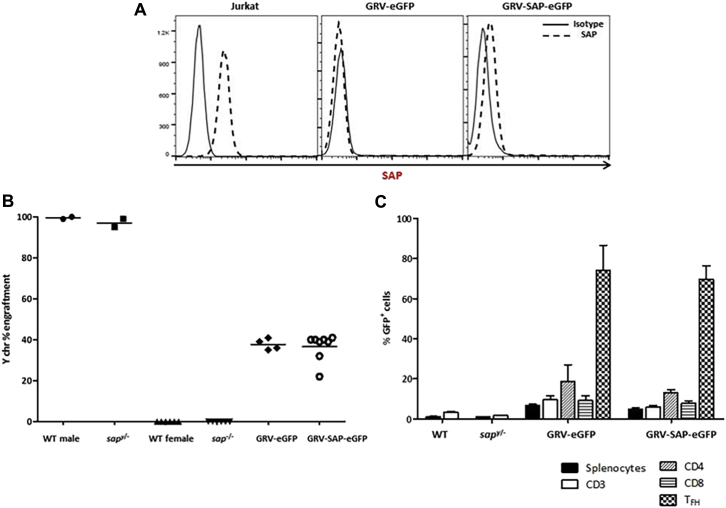
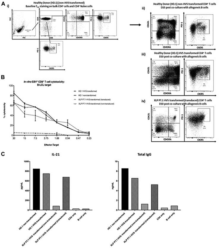
Methods: CD3+ lymphocytes from Sap-deficient mice were transduced with a gammaretroviral vector encoding human SAP cDNA before transfer into sublethally irradiated Sap-deficient recipients. After immunization with the T-dependent antigen 4-hydroxy-3-nitrophenylacetly chicken gammaglobulin (NP-CGG), recovery of humoral function was evaluated through germinal center formation and antigen-specific responses. To efficiently transduce CD3+ cells from patients, we generated an equivalent lentiviral SAP vector. Functional recovery was demonstrated by using in vitro cytotoxicity and T follicular helper cell function assays alongside tumor clearance in an in vivo lymphoblastoid cell line lymphoma xenograft model.

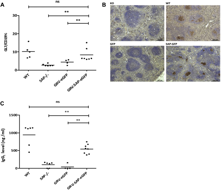
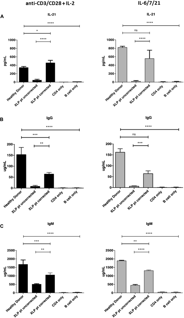
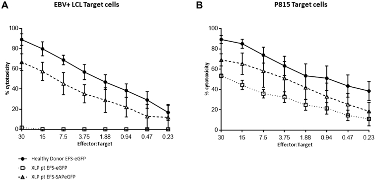
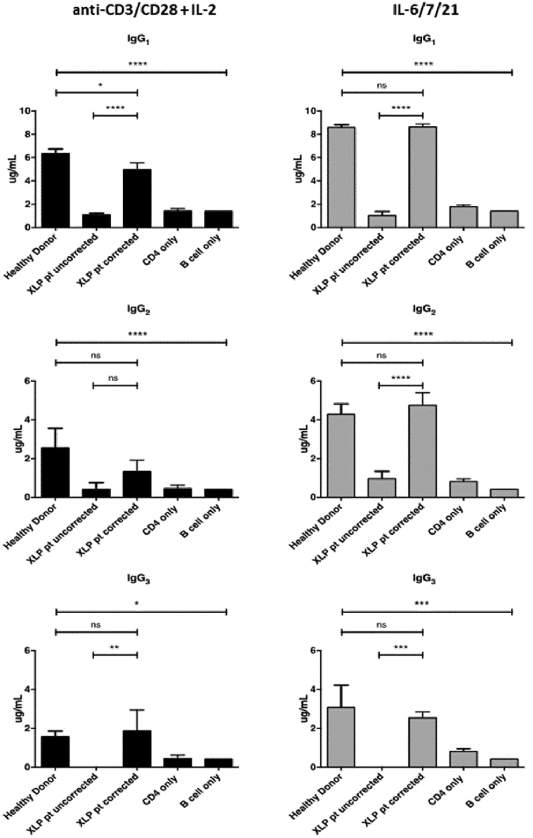
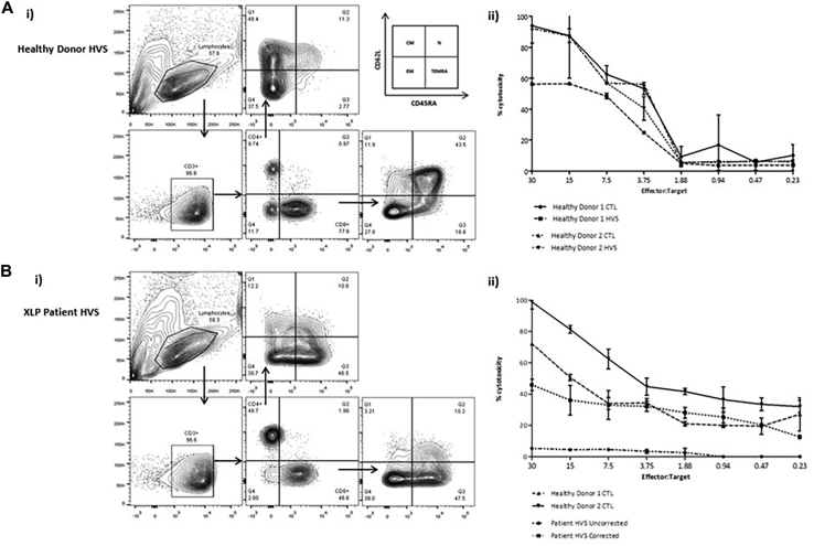
Results: In Sap-deficient mice 20% to 40% engraftment of gene-modified T cells led to significant recovery of germinal center formation and NP-specific antibody responses. Gene-corrected T cells from patients demonstrated improved cytotoxicity and T follicular helper cell function in vitro. Adoptive transfer of gene-corrected cytotoxic T lymphocytes from patients reduced tumor burden to a level comparable with that seen in healthy donor cytotoxic T lymphocytes in an in vivo lymphoma model.

Conclusions: These data demonstrate that autologous T-cell gene therapy corrects SAP-dependent defects and might offer an alternative therapeutic option for patients with X-linked lymphoproliferative disease 1.

Keywords: T-cell cytotoxicity; T-cell gene therapy; X-linked lymphoproliferative disease; follicular helper T cells.


Figures

Fig 1
Fig 1
Experimental design of the T-cell adoptive transfer model. A, Schematic representation of long terminal repeat (LTR)–driven gammaretroviral vector used for ex vivo transduction of murine donor splenic CD3+ selected T cells. i, Mock vector containing eGFP reporter gene only. ii, Vector containing codon-optimized SAP cDNA and eGFP. B, Timeline of adoptive T-cell transfer experiments after sublethal (6 Gy) irradiation at day 0 with intravenous infusion of transduced murine CD3+ T cells. Animals underwent tail vein bleeds at 3 and 6 weeks to assess peripheral T-cell donor engraftment before immunologic challenge at week 8 with the T cell–dependent antigen NP-CGG.
Fig 2
Fig 2
Analysis of humoral function after immunologic challenge. A, Analysis of germinal center B cells using flow cytometry in splenic lymphocytes stained with anti-CD19 and anti-GL7 antibodies. Results from individual mice are represented by dots, with mean values represented by a horizontal bar. B, Germinal center staining in splenic follicles using peanut agglutinin marking for germinal center B cells 10 days after immunization with NP-CGG at a magnification of ×40. Slides are representative of results seen in wild-type (WT) and Sap-deficient control animals and animals receiving gene-modified T cells (transduced with GRV-eGFP or GRV-SAP-eGFP vectors) demonstrating recovery of germinal centers in Sap-deficient mice receiving SAP-containing vector. C, NP-specific antibody production analysis with an ELISA performed on serum samples of all cohorts after NP-CGG vaccination, demonstrating functional restoration of germinal center activity in SAP-reconstituted animals comparable with that of wild-type littermates. **P < .05. ns, Not significant.
Fig 3
Fig 3
Correction of PBMC-derived TFH cells from patients with XLP1. A, Schematic representation of lentiviral vectors containing codon-optimized human SAP cDNA driven by the EFS promoter and eGFP or EFS-eGFP only. An MOI of 20 was used to transduce PBMC-derived selected naive CD4+ cells before in vitro B-cell coculture assay. Transduction efficiency ranged between 30% to 45% (data not shown), with a vector copy number of 2 to 3 copies per cell. B, Representative flow cytometric contour plots of the differentiated CD4+ cell phenotype 10 days after coculture with allogeneic tonsillar memory B cells or CD4 cell culture alone. Top 2 panels (middle to right), Healthy donor cells prestimulated with standard activation conditions (1.0 × 106 cells/mL) with a 1:1 anti-CD3/CD28 bead ratio plus human IL-2 (10 ng/mL) or the TFH cell–polarizing cytokines IL-6 (100 ng/mL), IL-7 (10 ng/mL), or IL-21 (20 ng/mL). Bottom 2 panels (left to right), PBMC-derived and HVS-transformed differentiated CD4+ T cells from patients with XLP1 by using the conditions as above. Cells were transduced 3 days after stimulation and cultured with or without allogeneic B cells. C, Recovery of the TFH cell population, as determined based on CXCR5 and PD-1 expression in corrected cells from patients with XLP1 after B-cell coculture assay in cells cultured in both anti-CD3/CD28/IL-2 and IL-6/IL-7/IL-21 culture conditions (n = 3). **P < .05. ns, Not significant.
Fig 4
Fig 4
Functional correction of TFH cells from patients with XLP1 in vitro. Quantification of IL-21 concentrations (A) and IgG (B) and IgM (C) levels in supernatants 10 days after coculture of naive CD4+ T cells and allogeneic B cells by means of ELISA. **P < .05, ***P < .001, and ****P < .0001. ns, Not significant.
Fig 5
Fig 5
SAP gene correction of CTLs from patients with XLP1 restores cytotoxicity in vitro. A,In vitro cytotoxic activity of CTLs generated from both healthy donors and EBV-seropositive patients with XLP1 (gene corrected and uncorrected) against allogeneic LCL target cells before intravenous infusion into NSG mice, as measured in a 51Cr release assay. Assays were performed in triplicates, and data shown are means ± SEMs of all values. B, Specificity of SAP function was determined by using non-LCL targets in a cytotoxicity assay in parallel. Murine mastocytoma P815 cells were cocultured with corrected and uncorrected effector cells from healthy donors and patients incubated with soluble anti-CD3. CTLS from all donors, including patients with XLP1, displayed cytotoxic activity, suggesting that EBV+ LCL-targeted killing is SAP and not CD3 mediated.
Fig 6
Fig 6
Phenotype of transduced and gene-corrected CTLs. A, Representative flow cytometric contour plots of CTL phenotype after in vitro stimulation with allogeneic LCLs in mock-transduced cells from healthy donors and patients and B, cells transduced with a corrective lentiviral SAP vector from patients. Transduction efficiency was assessed by using flow cytometry; eGFP expression ranged from 24% to 50%. CM, Central memory; EM, effector memory; N, naive; TEMRA, CD45RA+ effector memory. C, SAP expression from GFP+ selected uncorrected and gene-corrected cells from patients, as analyzed by using intracellular fluorescence-activate cell sorting staining. Solid line, Control IgG2b; dotted line, anti-SAP antibody.
Fig 7
Fig 7
Adoptive transfer of gene-corrected CTLs from patients with XLP1 induces regression of EBV+ LCL-generated tumors in an NSG mouse model. A, EBV+ B-LCL xenograft tumor model experimental design. B,i, Bioluminescence images of NSG mice 48 hours after subcutaneous LCL injections displaying formation of localized solid tumors. ii, Tumor burden after 10 days in untreated mice (top left panel, n = 3) and mice treated with healthy donor CTLs (3 donors, 3 animals per donor). iii, Tumor burden after 10 days in mice receiving uncorrected CTLs from patients with XLP1 (top panel) or gene-corrected CTLs from patients (bottom panel; 2 patient donors, 3 animals per donor) showing reduction in tumor burden in animals receiving gene-corrected cells from patients with XLP1. C, Dot plot representing quantification of tumor burden determined by using average photon density per second per square centimeter per steradian (p/s/cm2/sr) on the day of CTL infusions (D0), after 48 hours (D2), and after 10 days (D10). ***P < .0001.
Fig E1
Fig E1
Transduction efficiency and phenotype of murine splenic CD3+ donor T cells. A, Flow cytometric staining of murine T cells showing the gating strategy for CD4+ and CD8+ lymphocyte populations. B, Transduction efficiencies are determined by using GFP expression 72 hours after transduction with GRV-SAP-eGFP or GRV-eGFP alone. An average transduction of 50% to 60% is observed across T-lymphocyte compartments.
Fig E2
Fig E2
Analysis of adoptively transferred T cells. A, SAP protein expression was analyzed in gene-corrected murine splenic CD3+ T cells after reconstitution. B, Donor T-cell engraftment was assessed in whole splenocytes by using XY PCR techniques 10 weeks after reconstitution, showing average engraftment levels of 40%. C, Analysis of GFP levels in whole splenocytes and splenocyte-derived T-cell lineages in immunized animals.
Fig E3
Fig E3
Comparison of function between HVS-transformed cells and untransformed cells. One experiment was performed comparing phenotype (A), cytotoxicity (B), and TFH cell function (C) in HVS-transformed cells from healthy donors, HVS-transformed cells from patients, and nontransformed healthy donor cells, which demonstrated comparability among the all samples. No significant differences were observed in both cytotoxicity and TFH cells assays.
Fig E4
Fig E4
Functional correction of TFH cells from patients with XLP1 in vitro: IgG subclass production. Quantification of IgG subclass levels in supernatants 10 days after coculture of naive CD4+ T cells and allogeneic B cells by means of ELISA. **P < .05, ***P < .001, and ****P < .0001. ns, Not significant.
Fig E5
Fig E5
In vitro cytotoxicity of HVS-transformed CTLs by using an allogeneic LCL target. PBMCs from healthy donors and EBV+ patients with XLP were transformed with HVS. Ai and Bi, T-cell memory phenotyping was performed in both CD4 and CD8 populations to determine naive, CD45RA+ effector memory, effector memory, and central memory populations. Patient-transformed samples contained more terminally differentiated cells compared with healthy donors. Aii, 51Cr release assay demonstrated functional activity in HVS-transformed cells from healthy donors by using an allogeneic B-LCL target with no significant difference observed in cytotoxic activity between nontransformed and HVS-transformed cells. Bii, HVS-transformed cells from patients were transduced with the EFS-SAP-eGFP or EFS-eGFP mock vectors and subjected to a 51Cr release assay. Uncorrected patient samples showed no cytotoxic activity against allogeneic B-LCLs compared with patient-corrected CTLs, which were able to kill the LCL target within the range of 40% to 50%. No significant differences were observed between nontransformed healthy donor CTLs and patient-corrected HVS cells in cytotoxic activity when using an allogeneic B-LCL target.

Similar articles

Cited by

References

    1. Parolini S., Bottino C., Falco M., Augugliaro R., Giliani S., Franceschini R. X-linked lymphoproliferative disease. 2B4 molecules displaying inhibitory rather than activating function are responsible for the inability of natural killer cells to kill Epstein-Barr virus-infected cells. J Exp Med. 2000;192:337–346. - PMC - PubMed
    1. Tangye S.G., Phillips J.H., Lanier L.L., Nichols K.E. Functional requirement for SAP in 2B4-mediated activation of human natural killer cells as revealed by the X-linked lymphoproliferative syndrome. J Immunol. 2000;165:2932–2936. - PubMed
    1. Dupre L., Andolfi G., Tangye S.G., Clementi R., Locatelli F., Arico M. SAP controls the cytolytic activity of CD8+ T cells against EBV-infected cells. Blood. 2005;105:4383–4389. - PubMed
    1. Nakajima H., Cella M., Bouchon A., Grierson H.L., Lewis J., Duckett C.S. Patients with X-linked lymphoproliferative disease have a defect in 2B4 receptor-mediated NK cell cytotoxicity. Eur J Immunol. 2000;30:3309–3318. - PubMed
    1. Pasquier B., Yin L., Fondaneche M.C., Relouzat F., Bloch-Queyrat C., Lambert N. Defective NKT cell development in mice and humans lacking the adapter SAP, the X-linked lymphoproliferative syndrome gene product. J Exp Med. 2005;201:695–701. - PMC - PubMed

Publication types

MeSH terms

Substances